
Page 109 - Read Online
P. 109
Page 6 of 13 Bradley et al. Plast Aesthet Res 2019;6:11 I http://dx.doi.org/10.20517/2347-9264.2019.06
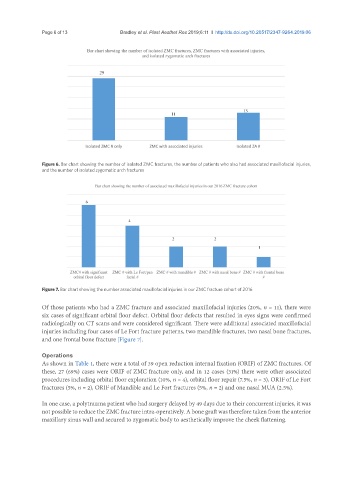
Figure 6. Bar chart showing the number of isolated ZMC fractures, the number of patients who also had associated maxillofacial injuries,
and the number of isolated zygomatic arch fractures
Figure 7. Bar chart showing the number associated maxillofacial injuries in our ZMC fracture cohort of 2016
Of those patients who had a ZMC fracture and associated maxillofacial injuries (20%, n = 11), there were
six cases of significant orbital floor defect. Orbital floor defects that resulted in eyes signs were confirmed
radiologically on CT scans and were considered significant. There were additional associated maxillofacial
injuries including four cases of Le Fort fracture patterns, two mandible fractures, two nasal bone fractures,
and one frontal bone fracture [Figure 7].
Operations
As shown in Table 1, there were a total of 39 open reduction internal fixation (ORIF) of ZMC fractures. Of
these, 27 (69%) cases were ORIF of ZMC fracture only, and in 12 cases (31%) there were other associated
procedures including orbital floor exploration (10%, n = 4), orbital floor repair (7.5%, n = 3), ORIF of Le Fort
fractures (5%, n = 2), ORIF of Mandible and Le Fort fractures (5%, n = 2) and one nasal MUA (2.5%).
In one case, a polytrauma patient who had surgery delayed by 49 days due to their concurrent injuries, it was
not possible to reduce the ZMC fracture intra-operatively. A bone graft was therefore taken from the anterior
maxillary sinus wall and secured to zygomatic body to aesthetically improve the cheek flattening.