
Page 240 - Read Online
P. 240
Khan et al. Modified lower eyelid blepharoplasty
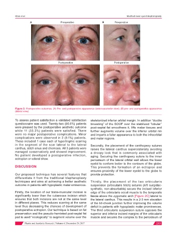
A Preoperative B Preoperative
Postoperative Postoperative
Figure 5: Postoperative outcomes. (A) Pre- and postoperative appearance (antero-posterior view); (B) pre- and postoperative appearance
(lateral view)
To assess patient satisfaction a validated satisfaction skeletonized inferior orbital margin. In addition “double
questionnaire was used. Twenty-two (66.6%) patients breasting” of the SOOF over the stabilized “lobular”
were pleased by the postoperative aesthetic outcome post-septal fat smoothens it, lifts malar tissues and
while 11 (33.3%) patients were satisfied. There further augments volume over the inferior orbital rim
were no major postoperative complications. Minor and imparts a fuller appearance to both the infraorbital
complications were observed in 3 (9.1%) patients. and malar regions.
These included 1 case each of hypertrophic scarring
in the segment of the scar lateral to the lateral Secondly, the placement of the canthopexy sutures
canthus, stitch sinus and chemosis. All 3 patients were raises the lateral canthus superolaterally avoiding
managed conservatively and showed improvement. a droopy look that is commonly associated with
No patient developed a postoperative infection, aging. Securing the canthopexy suture to the inner
ectropion or scleral show. periosteum of the lateral orbital wall allows the lower
eyelid to conform better to the contours of the globe.
DISCUSSION This prevents the formation of an ectropion and
ensures proximity of the lower eyelid to the globe to
Our proposed technique has several features that provide protection.
differentiate it from the traditional blepharoplasty
techniques and aims at achieving a better aesthetic Thirdly, the placement of the two orbicularis
outcome in patients with hypoplastic malar eminences. suspension (orbicularis hitch) sutures (4/0 surgidac-
synthetic, non-absorbable) secure the incised inferior
Firstly, the location of our trans-muscular incision is edge of the orbicularis occuli muscle to the temporalis
significantly lower than the cutaneous incision which fascia above the zygomatic arch [Figure 2C] lateral to
ensures that both incisions are not at the same level the lateral canthus. This results in a 2-3 mm elevation
in different planes. This reduces scarring at the same of the lid-cheek junction further improving the volume
level thus decreasing the chances of development of deficit in patients with hypoplastic malar prominences.
postoperative ectropion. Our technique is based on fat The third orbicularis suspension suture opposes the
preservation and the pseudo-herniated post-septal fat superior and inferior incised margins of the orbicularis
pad is used “ecologically” to augment volume over the muscle and secures the complex to the periosteum of
Plastic and Aesthetic Research ¦ Volume 4 ¦ December 29, 2017 233