
Page 238 - Read Online
P. 238
Khan et al. Modified lower eyelid blepharoplasty
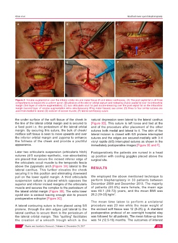
A B C
D E F
Figure 2: Volume augmentation over the inferior orbital rim and malar tissue lift and lateral canthopexy. (A) The post septal fat in all three
compartments is teased into a uniform apron; (B) plication of the inferior orbital septum and redraping of post septal fat over the infraorbital
margin (first layer of volume augmentation); (C) sub orbicularis oculi fat pad double-breasting over the post septal fat on the infraorbital
margin (second layer of volume augmentation while simultaneously lifting malar tissues) see arrow; (D) three to four similar sutures are
used from medial to lateral; (E) excision of excess muscle; (F) lateral canthopexy suture
the under surface of the soft tissue of the cheek in natural depression seen lateral to the lateral canthus
the line of the lateral orbital margin and is secured to [Figure 3D]. This suture is left loose and tied at the
a fixed point i.e. the periosteum of the lateral orbital end of the procedure after placement of the other
margin. By securing this suture, the bulk of cheek/ sutures both medal and lateral to it. The skin of the
midface soft tissue is seen to move upwards and over lateral incision is closed with 6/0 prolene interrupted
the inferior orbital margin and zygoma to enhance sutures and the edges are secured medially with 3-4
the fullness of the cheek and provide a youthful vicryl rapide (6/0) interrupted sutures as shown in the
appearance. immediately postoperative images [Figure 3E and F].
Later two orbicularis suspension (orbicularis hitch) Postoperatively the patients are nursed in a head
sutures (4/0 surgidac-synthetic, non-absorbable) up position with cooling goggles placed above the
are placed that secure the incised inferior edge of surgical site.
the orbicularis occuli muscle to the temporalis fascia
above the zygomatic arch [Figure 3A] lateral to the RESULTS
lateral canthus. This further elevates the cheek
securing it in this position and eliminating downward
pull on the lower eyelid margin. A third orbicularis We employed the above mentioned technique to
suspension suture is placed which opposes the perform blepharoplasty in 33 patients between
superior and inferior incised margins of the orbicularis December 2009 and December 2013. The majority
muscle and secures the complex to the periosteum of of patients (60.6%) were female, the mean age
the lateral orbital margin [Figure 3B]. The extra lower was 49.1 (39-72) years, and the mean BMI was
2
eyelid skin is excised leaving enough skin to prevent 26.2 (19-33) kg/m .
postoperative ectropion [Figure 3C].
The mean time taken to perform a unilateral
A lateral contouring suture is then placed using 5/0 procedure was 23 min while the mean weight of
prolene, through the skin edges just lateral to the the excised soft tissue was 12 (8-25) g. A standard
lateral canthus to secure them to the periosteum of postoperative protocol of an overnight hospital stay
the lateral orbital margin. This “quilting” facilitates was followed for all patients. The mean follow-up time
the creation of a smooth trough which is the was 14 (12.5-19) months. The outcomes of interest
Plastic and Aesthetic Research ¦ Volume 4 ¦ December 29, 2017 231