
Page 237 - Read Online
P. 237
Khan et al. Modified lower eyelid blepharoplasty
A B C
D E F
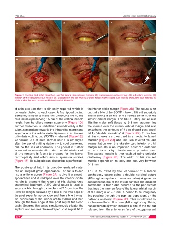
Figure 1: Incision and initial dissection. (A) The lateral skin incision marking; (B) subcutaneous undermining; (C) sub-ciliary incision; (D)
incision of the orbicularis oculi muscle; (E) dissection in the sub-muscular plane releasing the fascia over the sub orbicularis oculi fat pad; (F)
orbito-malar ligament release and lateral pocket dissection
of skin excision that is clinically required which is the inferior orbital margin [Figure 2B]. The suture is not
generally titrated to each case. A fine tipped cutting cut and a bite of the SOOF is taken, lifting it superiorly
diathermy is used to incise the underlying orbicularis and securing it on top of the redraped fat over the
oculi muscle preserving 1.5 cm of the vertical muscle inferior orbital margin. This SOOF lifting suture also
height from the ciliary margin superiorly [Figure 1D]. lifts the malar soft tissue by 2-3 mm, augmenting
Further dissection is undertaken infero-laterally in the the volume over the inferior orbital margin and also
submuscular plane towards the infraorbital margin and smoothens the contours of the re-draped post septal
zygoma and the orbito-malar ligament over the sub fat by “double breasting” it [Figure 2C]. Three-four
orbicularis oculi fat pad (SOOF) is released [Figure 1E]. similar sutures are then used in a medial to lateral
Generous use of cold normal saline is employed manner [Figure 2D] and this two-layered volume
after the use of cutting diathermy to cool tissue and augmentation over the skeletonized inferior orbital
reduce the risk of chemosis. The pocket is further margin results in an improved aesthetic outcome
extended supero-laterally under the orbicularis oculi in patients with hypolastic malar prominences.
till the temporalis fascia to prepare for the lateral The excess muscle is then excised using unipolar
canthoplasty and orbicularis suspension sutures diathermy [Figure 2E]. The width of this excised
[Figure 1F]. No subperiosteal dissection is performed. muscle depends on its laxity and can vary between
5-12 mm.
The post-septal fat, in its pseudo-herniated state,
has an irregular gross appearance. The fat is teased This is followed by the placement of a lateral
into a uniform apron [Figure 2A] to give it a smooth canthopexy suture using a double needled suture
appearance and is redraped over the inferior orbital (4/0 surgidac-synthetic, non-absorbable). A generous
margin to augment the volume of this skeletonized subcutaneous bite of the lateral canthus and adjacent
anatomical landmark. A 5/0 vicryl suture is used to soft tissue is taken and secured to the periosteum
secure a bite through the septum at 2.5 cm from the that lines the inner surface of the lateral orbital margin
lower lid margin, followed by a bite of the free edge of at the margin or 2-3 mm superior to an imaginary
the post septal fat apron edge and then a bite through line passing through the pupil as dependent on the
the periosteum of the inferior orbital margin and then patient’s anatomy [Figure 2F]. This is followed by
through the free edge of the post septal fat apron a cheek/midface lift suture (4/0 surgidac-synthetic,
again. Securing this suture simultaneously plicates the non-absorbable) which includes a bite of the fat pad
septum and secures the re-draped post septal fat to released from the anterior surface of the zygoma and
230 Plastic and Aesthetic Research ¦ Volume 4 ¦ December 29, 2017