
Page 236 - Read Online
P. 236
Khan et al. Modified lower eyelid blepharoplasty
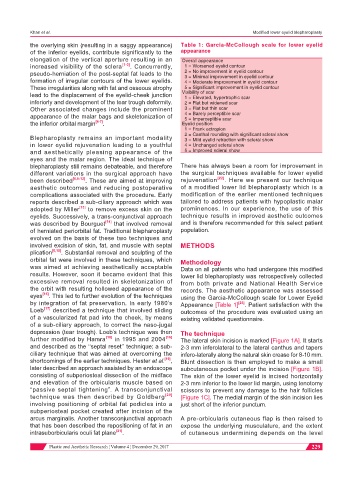
the overlying skin (resulting in a saggy appearance) Table 1: Garcia-McCollough scale for lower eyelid
of the inferior eyelids, contribute significantly to the appearance
elongation of the vertical aperture resulting in an Overall appearance
increased visibility of the sclera [1-3] . Concurrently, 1 = Worsened eyelid contour
pseudo-herniation of the post-septal fat leads to the 2 = No improvement in eyelid contour
3 = Minimal improvement in eyelid contour
formation of irregular contours of the lower eyelids. 4 = Moderate improvement in eyelid contour
These irregularities along with fat and osseous atrophy 5 = Significant improvement in eyelid contour
lead to the displacement of the eyelid-cheek junction Visibility of scar
1 = Elevated, hypertrophic scar
inferiorly and development of the tear trough deformity. 2 = Flat but widened scar
Other associated changes include the prominent 3 = Flat but thin scar
appearance of the malar bags and skeletonization of 4 = Barely perceptible scar
5 = Imperceptible scar
the inferior orbital margin [4-7] . Eyelid position
1 = Frank ectropion
Blepharoplasty remains an important modality 2 = Canthal rounding with significant scleral show
3 = Mild eyelid retraction with scleral show
in lower eyelid rejuvenation leading to a youthful 4 = Unchanged scleral show
and aesthetically pleasing appearance of the 5 = Improved scleral show
eyes and the malar region. The ideal technique of
blepharoplasty still remains debateable, and therefore There has always been a room for improvement in
different variations in the surgical approach have the surgical techniques available for lower eyelid
been described [6,8-12] . These are aimed at improving rejuvenation [22] . Here we present our technique
aesthetic outcomes and reducing postoperative of a modified lower lid blepharoplasty which is a
complications associated with the procedure. Early modification of the earlier mentioned techniques
reports described a sub-ciliary approach which was tailored to address patients with hypoplastic malar
adopted by Miller [13] to remove excess skin on the prominences. In our experience, the use of this
eyelids. Successively, a trans-conjunctival approach technique results in improved aesthetic outcomes
was described by Bourguet [14] that involved removal and is therefore recommended for this select patient
of herniated periorbital fat. Traditional blepharoplasty population.
evolved on the basis of these two techniques and
involved excision of skin, fat, and muscle with septal METHODS
plication [6,15] . Substantial removal and sculpting of the
orbital fat were involved in these techniques, which Methodology
was aimed at achieving aesthetically acceptable Data on all patients who had undergone this modified
results. However, soon it became evident that this lower lid blepharoplasty was retrospectively collected
excessive removal resulted in skeletonization of from both private and National Health Service
the orbit with resulting hollowed appearance of the records. The aesthetic appearance was assessed
eyes [16] . This led to further evolution of the techniques using the Garcia-McCollough scale for Lower Eyelid
by integration of fat preservation. In early 1980’s Appearance [Table 1] [23] . Patient satisfaction with the
Loeb [17] described a technique that involved sliding outcomes of the procedure was evaluated using an
of a vascularized fat pad into the cheek, by means existing validated questionnaire.
of a sub-ciliary approach, to correct the naso-jugal
depression (tear trough). Loeb’s technique was then The technique
further modified by Hamra [18] in 1995 and 2004 [16] The lateral skin incision is marked [Figure 1A]. It starts
and described as the ‘‘septal reset’’ technique; a sub- 2-3 mm inferolateral to the lateral canthus and tapers
ciliary technique that was aimed at overcoming the infero-laterally along the natural skin crease for 8-10 mm.
shortcomings of the earlier techniques. Hester et al. [19] , Blunt dissection is then employed to make a small
later described an approach assisted by an endoscope subcutaneous pocket under the incision [Figure 1B].
consisting of subperiosteal dissection of the midface The skin of the lower eyelid is incised horizontally
and elevation of the orbicularis muscle based on 2-3 mm inferior to the lower lid margin, using tenotomy
“passive septal tightening”. A transconjunctival scissors to prevent any damage to the hair follicles
technique was then described by Goldberg [20] [Figure 1C]. The medial margin of the skin incision lies
involving positioning of orbital fat pedicles into a just short of the inferior punctum.
subperiosteal pocket created after incision of the
arcus marginalis. Another transconjunctival approach A pre-orbicularis cutaneous flap is then raised to
that has been described the repositioning of fat in an expose the underlying musculature, and the extent
intrasuborbicularis oculi fat plane [21] . of cutaneous undermining depends on the level
Plastic and Aesthetic Research ¦ Volume 4 ¦ December 29, 2017 229