
Page 11 - Read Online
P. 11
Fok et al. Skin temperature of laser for Nevus of Ota
Table 1: Skin surface temperatures for Q-switched ruby Light dosage
2
laser treatment for different energy density 55 (J/cm )
Skin surface temperature (℃ ) † 50 6
Energy density (J/cm ) 45 7
2
IR* Thermal wave equation ) 8
6 40.8 ± 0.3 41.2 ± 0.2 Skin surface temperature (℃ 40
7 41.9 ± 0.3 42.5 ± 0.4 35 9
8 44.1 ± 0.4 44.3 ± 0.3 30 10
9 46.9 ± 0.2 47.6 ± 0.2 0 1 2 3 4 5 6 7 8 9 10 11 12 13 14 15 Control
10 50.8 ± 0.4 51.5 ± 0.4 Time (s)
†
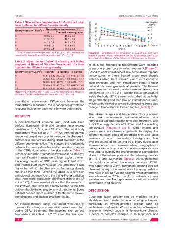
: baseline skin surface temperature -32.4 ± 0.2 ℃ ; IR*: temperatures Figure 3: Temperature determination of superficial skin with
measured by infrared thermal image instrument. P = 2 × 10 -11 infrared thermal image instrument for the Q-switched ruby laser
treatment of the Nevus of Ota patients in different energy density
Table 2: Mean melanin index of clearing and fading
response of Nevus of Ota after Q-switched ruby laser of 15 s, the changes in temperature were recorded
treatment for different energy density to assume proper care following treatment [Figure 3].
2
Energy density (J/cm ) 1 month 3 months 6 months Based on what was observed in superficial skin regions,
6 61.81 ± 1.42 55.77 ± 1.76 54.57 ± 1.75 temperatures in those treated areas rose sharply
7 55.13 ± 1.57 54.82 ± 1.63 53.68 ± 1.47 within 5 s when there was a “T-jump” in response to
8 52.23 ± 1.08 47.85 ± 1.39 44.76 ± 1.11 laser exposure, and then immediately began to taper
9 51.46 ± 1.28 46.69 ± 1.07 40.47 ± 1.46 out and decrease gradually afterwards. The thermal
10 49.62 ± 1.54 45.78 ± 1.74 40.19 ± 1.53 wave equation showed that the baseline skin surface
Mean-index of normal skin = 32.21 ± 2.75; mean-index of Nevus of temperature (32.4 ± 0.2 ℃) and the tissue temperature
Ota before Q-switched ruby laser treatment = 63.15 ± 1.44
inside the body (37 ℃) were undisturbed at the initial
quantitative assessment. Differences between the stage of heating and then took an instantaneous jump,
temperatures measured and clearing/depigmentation which can be viewed as a wave front resulting from a step
[31]
response indices for each test site were determined. change in temperature at the skin surface [Table 1].
The infrared images and temperature plots of normal
RESULTS skin and oculodermal melanosis-affected skin
A one-dimensional equation was used with fixed represent a patient’s reaction time post-treatment, with
2
surface illumination time and variable laser energy a QSRL energy density of 9 J/cm and the resulting
densities of 6, 7, 8, 9, and 10 J/cm . The initial body temperature [Figures 4 and 5]. Other images and
2
temperature was set at 37 ℃. [30] An infrared thermal graphs were also taken of patients to display the
different reaction times of superficial skin after laser
image instrument was used to measure the changes in treatment, in which temperature averages are clear
surface skin temperature during QSRL treatment at the over the course of 10, 20, and 30 s. Injury due to laser
different energy densities. This showed the relationship illumination can be minimized while using optimum
between the energy densities and temperature changes dosage to treat Nevus of Ota. A dermospectrometer
of the QSRL illumination of the skin surface [Table 1]. was used to quantify the improvement in pigmentation
Temperatures in the treated areas were observed to have at each of the follow-up visits at the following intervals
risen significantly in response to laser exposure when of 1, 3, 6, and 12 months [Table 2]. Although thermal
the energy density of QSRL was higher than 8 J/cm burns did occur when the energy density of QSRL
2
and thermal burn injury resulted (the temperature was was higher than 8 J/cm , permanent scarring was not
2
higher than 44 ℃). In other words, the energy density observed on any of the treated sites. Hyperpigmentation
should be less than 8 J/cm if the QSRL is to treat skin was noted in 5% (n = 2) and delayed hypopigmentation
2
pathological changes. Using the Irving-Fisher statistical was observed in 2.5% (n = 1) of patients but was
test, there were statistically significant differences (P transient and resolved spontaneously without medical
= 2 × 10 ). These results indicate that the severity of intervention in all patients.
-11
the involved area was not directly related to the final
outcome but to the energy density of treatments. Some DISCUSSION
patients needed more number of treatment to prevent
complications and sustain excellent results. Cutaneous laser surgery can be modeled on the
short-term heat transfer behavior of iological tissues,
An infrared thermal image instrument was used to particularly in hyperpigmented lesions such as
measure the changes in superficial skin temperature oculodermal melanosis. When the surface of biological
during QSRL treatment. The baseline skin surface tissues is heated causing a temperature change,
temperature was 32.4 ± 0.2 ℃. Over the time span a series of complex changes in its biophysics and
4 Plastic and Aesthetic Research ¦ Volume 4 ¦ January 19, 2017