
Page 265 - Read Online
P. 265
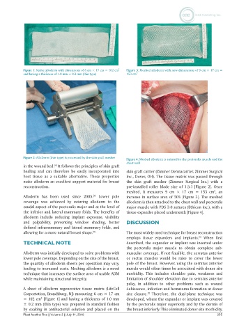
Figure 1: Native alloderm with dimensions of 6 cm × 17 cm = 102 cm   2  Figure 3: Meshed alloderm with new dimensions of 9 cm × 17 cm =
            and having a thickness of 1.0 mm ± 0.2 mm (thin type)  153 cm 2


            Figure 2: AlloDerm (thin-type) is processed by the skin graft mesher  Figure 4: Meshed alloderm is sutured to the pectoralis muscle and the
                                                              chest wall
            in the wound bed.  It follows the principles of skin graft
                           [4]
            healing and  can  therefore be easily incorporated  into   skin graft carrier (Zimmer Dermacarrier, Zimmer Surgical
            host tissue  as a suitable  alternative.  These  properties   Inc., Dover, OH). The tissue matrix was passed through
            make alloderm an excellent support material for breast   the skin graft mesher  (Zimmer Surgical Inc.) with a
            reconstruction.                                   pre-installed roller blade size  of 1.5:1 [Figure 2]. Once
                                                              meshed, it measures 9 cm × 17  cm = 153  cm , an
                                                                                                         2
            Alloderm has been used since 2005.  Lower pole    increase in surface area of 50% [Figure 3]. The meshed
                                              [5]
            coverage was achieved by  suturing  alloderm to the   alloderm is then attached to the chest wall and pectoralis
            caudal aspect of the pectoralis major and at the level of   major muscle with PDS 2-0 sutures (Ethicon Inc.), with a
            the inferior and lateral mammary folds. The benefits of   tissue expander placed underneath [Figure 4].
            alloderm include reducing implant exposure,  visibility
            and palpability, preventing window shading, better   DISCUSSION
            defined inframammary and lateral mammary folds, and
            allowing for a more natural breast shape. [5]     The most widely used technique for breast reconstruction
                                                              employs tissue  expanders and implants.  When first
                                                                                                 [6]
            TECHNICAL NOTE                                    described, the expander or implant was inserted under
                                                              the pectoralis major muscle to obtain complete sub-
            AlloDerm was initially developed to solve problems with   muscular coverage. If not feasible, the serratus anterior
            lower pole coverage. Depending on the size of the breast,   or rectus muscles would be raise to cover the lower
            the quantity of alloderm sheets per operation may vary,   pole of the breast. However, using the serratus anterior
            leading to increased costs. Meshing alloderm is a novel   muscle would often times be associated with donor site
            technique that increases the surface area of usable ADM   morbidity.  This  includes shoulder pain, weakness and
            while maintaining structural integrity.           limitation of shoulder elevation due to serratus anterior
                                                              palsy, in addition to other problems such as wound
            A sheet of alloderm regenerative tissue matrix (LifeCell   dehiscence, infection and hematoma formation at donor
            Corportation, Branchburg, NJ) measuring 6 cm × 17 cm   site closure.  Therefore, the dual-plane technique was
                                                                        [7]
                     2
            = 102 cm  [Figure 1] and having a thickness of 1.0 mm   developed, where the expander or implant was covered
            ± 0.2 mm (thin type) was prepared in standard fashion   by the pectoralis major superiorly and by the dermis of
            by soaking in antibacterial solution and placed on  the   the breast inferiorly. This eliminated donor site morbidity,
            Plast Aesthet Res || Volume 3 || July 14, 2016                                                255