
Page 137 - Read Online
P. 137
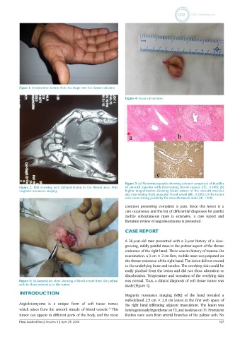
Figure 1: Preoperative picture. Note the bulge over the thenar eminence

                                                               Figure 4: Gross cut section


                                                               Figure 5: (a) Photomicrographs showing a tumor composed of bundles
           Figure 2: MRI showing well defined lesion in the thenar area. MRI:   of smooth muscles with intervening blood vessels (HE, ×100); (b)
           magnetic resonance imaging                          higher magnification showing bland nature of the smooth muscles
                                                               and intervening thick muscular blood vessel (HE, ×200); (c) the tumor
                                                               cells show strong positivity for smooth muscle actin (IP, ×100)
                                                               common  presenting  complaint  is  pain.  Since  this  lesion  is  a
                                                               rare occurrence and the list of differential diagnoses for painful
                                                               mobile subcutaneous mass is extensive, a case report and
                                                               literature review of angioleiomyoma is presented.

                                                               CASE REPORT

                                                               A 36-year-old man presented with a 2-year history of a slow-
                                                               growing, mildly painful mass in the palmar aspect of the thenar
                                                               eminence of the right hand. There was no history of trauma. On
                                                               examination, a 3 cm × 2 cm firm, mobile mass was palpated on
                                                               the thenar eminence of the right hand. The lesion did not extend
                                                               to the underlying bone and tendon. The overlying skin could be
                                                               easily pinched from the lesion and did not show ulceration or
                                                               discoloration. Temperature and sensation of the overlying skin
           Figure 3: Intraoperative view showing a blood vessel from the palmar   was normal. Thus, a clinical diagnosis of soft tissue tumor was
           arch in close proximity to the tumor                made [Figure 1].
           INTRODUCTION                                        Magnetic resonance imaging (MRI) of the hand revealed a
                                                               well-defined 2.5 cm × 2.8 cm lesion in the first web space of
           Angioleiomyoma is  a unique  form  of soft tissue tumor,   the right hand infiltrating adjacent musculature. The lesion was
           which arises from the smooth muscle of blood vessels.  This   heterogeneously hyperdense on T2, and isodense on T1. Prominent
                                                       [1]
           tumor can appear in different parts of the body, and the most   feeders were seen from arterial branches of the palmar arch. No
           Plast Aesthet Res || Volume 3 || April 25, 2016                                                    127