
Page 129 - Read Online
P. 129
and this highlight the wide range of patient age group that has been documented with minimal complications. However, it is
can be successfully reconstructed using this flap. Healing is recommended that early flap division should not be undertaken
usually excellent in children following use of forehead flap in active smokers and in patients with bleeding disorders to
and this has been attributed to the non sebaceous quality of avoid complications. [21,27]
their forehead skin. [15]
Documented disadvantages of the forehead flap include
Trauma (mainly road traffic crash) was the main aetiological facial disfiguring and bulkiness of flap. Complications noted
factor for orofacial defect, followed by neoplasia. Delayed in this study are shown in Table 3. Infective complications
reconstruction was used in most patients and this may be related were observed only in patients who were reconstructed using
to the aetiological factors. Most road traffic crash soft tissue complete forehead flap. This increased tendency for infection
injuries in our environment present as class III or IV surgical with complete forehead flap may be related to the large surface
wounds and require meticulous wound care to become clean area of the flap exposed.
before reconstruction can be undertaken. This fact has been
highlighted in studies from this environment. [16,17] Total flap failure was recorded in 2 cases (1 complete and
1 partial forehead flap). Failure of the median forehead flap
Complete forehead flap was the most common type of flap used, occurred post division despite a timing period of 36 days prior
accounting for 72.1% of all forehead flaps in our study. This is in to division. It is likely that excessive pressure was applied to
contrast to other studies [18,19] that reported partial forehead flaps the distal part of the flap during division or the patient had
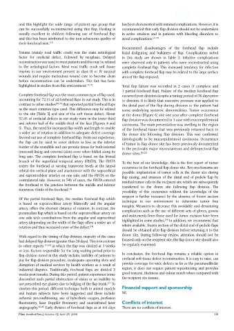
as the most common type used. This difference may be related some underlying systemic abnormalities. Tumor occurrence
to the site [Table 3] and size of the soft tissue defect. About at the donor [Figure 6] site one year after complete forehead
57.4% of orofacial defects in our study were in the lower third flap division was documented in 1 case with mucoepidermoid
and inferior half of the middle-third of the face [Figures 4 and carcinoma. The main presentation was swelling in the region
5]. Thus, the need for increased flap width and length to enable of the forehead tissue that was previously returned back to
a wider arc of rotation in addition to adequate defect coverage the donor site following flap division. This was confirmed
favored our use of complete forehead flap. From our experience, histologically to be mucoepidermoid carcinoma. Occurrence
the flap can be used to cover defects as low as the inferior of tumor in flap donor site has been previously documented
border of the mandible and can provide tissue for both internal in the pectoralis major myocutaneous and deltopectoral flap
(mucosal) lining and external (skin) cover when folded along its donor sites. [28,29]
long axis. The complete forehead flap is based on the frontal
branch of the superficial temporal artery (FBSTA). The FBSTA To the best of our knowledge, this is the first report of tumor
enters the forehead at varying transverse levels at the lateral occurrence in the forehead flap donor site. Two mechanisms are
orbital rim vertical plane and anastomose with the supraorbital possible: implantation of tumor cells in the donor site during
and supratrochlear arteries on one side, and the FBSTA on the flap raising, and invasion of the distal end of pedicle flap by
contralateral side. However, in 74% of cases, the FBSTA entered residual tumor cells in the recipient site which are subsequently
the forehead at the junction between the middle and inferior transferred to the donor site following flap division. The
transverse thirds of the forehead. [14]
possibility of this occurrence without the knowledge of the
surgeon is further increased by the absence of frozen section
Of the partial forehead flaps, the median forehead flap which technique in our environment to determine tumor free
is based on supratrochlear artery bilaterally and the angular margins. Measures to decrease this avoidable and devastating
artery, offers the shortest distance of rotation. In contrast, the complication such as the use of different sets of gloves, gowns
paramedian flap which is based on the supratrochlear artery on and instruments from those used for tumor excision have been
one side with contributions from the angular and supraorbital [30]
artery (depending on the width of the flap) offers a wider arc of highlighted in some studies. In addition, we recommend that
rotation and thus increased cover of the defect. [14] where available, frozen section of the distal end of pedicle flaps
should be obtained after flap division before returning it to the
With regard to the timing of flap division, majority of the cases donor site. During follow-up review, attention should not be
had delayed flap division (greater than 28 days). This is in contrast focused only on the recipient site; the flap donor site should also
to other reports [13,20] in which the flap was divided at 3 weeks be regularly examined.
or less. Factors responsible for the long waiting period prior to
flap division noted in this study include; inability of patients to In conclusion, the forehead flap remains a reliable option in
pay for flap division procedure, inadequate operating slots and orofacial soft tissue defect reconstruction. It is easy to raise, can
disruption of medical services by health workers as a result of provide coverage for wide defects as far as the paramandibular
industrial disputes. Traditionally, forehead flaps are divided 3 region, it does not require patient repositioning and provides
weeks post transfer. During this period, patient experience some good textural, thickness and colour match when compared with
discomfort such partial obstruction of vision or an inability to the recipient site tissues.
[21]
use prescribed eye glasses due to bulging of the flap trunk. To
shorten this period, different technique both in animal models Financial support and sponsorship
and human subjects have been suggested and these include Nil.
ischemic preconditioning, use of hyberbaric oxygen, perfusion
fluorometry, laser Doppler flowmetry and near-infrared laser Conflicts of interest
angiography. [22-26] Early division of forehead flaps as at 4-6 days There are no conflicts of interest.
Plast Aesthet Res || Volume 3 || April 25, 2016 119