Page 364 - Read Online
P. 364
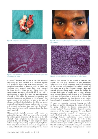
Figure 5: Complete excision specimen                Figure 6: Postoperative photograph after complete healing (three months
                                                              after surgery)


          Figure  7: Hematoxylin and eosin stain shows variable size spores with
          sporangia in the lumen                              Figure 8: Periodic acid‑Schiff stain highlighting the wall of spores

          R.  seeberi.  Recently, an analysis of the 18S ribosomal   canthus.  The reasons  for  the  spread  of  infection  are
                  [1]
          ribonucleic acid  gene  classified it  as  a protistan  parasite   unclear, and may occur secondary to local inoculation
          and included it  in the new class  mesomycetozoea.  The   following trauma which progresses with local  replication
                                                     [2]
          organism  is  prevalent  in  Southern  India,  Sri  Lanka  and   of the organism and associated hyperplastic growth of
          Southeast  Asia,  although cases  have  been  reported   host tissue and a localized immune response. Nasal and
          in  South  America,  Africa and the  United States.  The   mucosal rhinosporidiosis usually spread by bathing in
          most  common  age  group affected is  15‑40  years  with  a   stagnant  fresh  water  of  ponds,  lakes  or  rivers  whereas
          predominance in males. The nose and  nasopharynx are   ocular rhinosporidiosis spreads by dust or air.  In cases in
                                                                                                    [5]
          affected in 70%, while the palpebral conjunctivae and   which  only  the  lacrimal  sac  is  involved,  it  is  believed  that
          associated structures  are affected in 15% of cases.  The   the infection reaches the sac from the nose or eye via the
                                                     [3]
          mouth,  upper airway  and eye  may  also be  the  sites  of   lacrimal canaliculi without affecting the nose or conjunctiva.
          disease. Additional sites including the skin, ear, larynx,   CT  scan  and  magnetic  resonance  imaging  can  help
          trachea, bronchi, genitals (vagina, penile urethra or meatus   in diagnosis and in determining the extent of disease
          and scrotum),  and rectum  have  also been  described. [4,5]    by giving moderate to intense enhancement in
          Deep  organ  involvement  with  systemic  disease  has  been   contrast  studies.   However,  definitive  diagnosis  requires
                                                                             [6]
          rarely reported.
                                                              microscopy  of the biopsy specimen. The oval‑shaped
          Rhinosporidiosis typically presents with sessile or   sporangia,  containing  hundreds  of  endospores,  are
          pedunculated  polyps,  which  are  occasionally  surrounded   easily identified under the microscope. Fungal stains
          by  whitish  spores.  Symptoms  will  depend  upon  the  site   including  Gomori  methenamine  silver,  periodic
          affected. Nasal symptoms include nasal obstruction,   acid‑Schiff  and potassium chloride  also help  in  the
          postnasal drip and bleeding. Ocular symptoms include   diagnosis.  Serological  tests such as the enzyme‑linked
                                                                                                              [7]
          redness of conjunctiva, itching, epiphora and photophobia.   immunosorbent assay are used for epidemiological studies.
          When only the lacrimal sac is affected, it may present as   The differential diagnosis includes condyloma accuminata
          a  small,  isolated  and  nontender  swelling  over  the  medial   and hemangioma. Although rhinosporidiosis cannot


          Plast Aesthet Res || Vol 2 || Issue 6 || Nov 12, 2015                                             355

