Page 363 - Read Online
P. 363
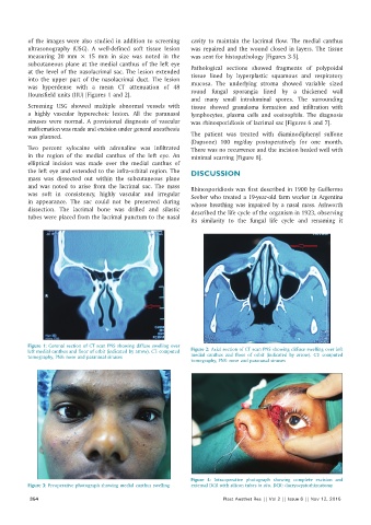
of the images were also studied in addition to screening   cavity to maintain  the  lacrimal flow.  The  medial  canthus
          ultrasonography  (USG). A  well‑defined soft tissue  lesion   was repaired and the wound closed in layers. The tissue
          measuring  20  mm  ×  15  mm in size  was noted in the   was sent for histopathology [Figures 3‑5].
          subcutaneous plane at the medial canthus of the left eye   Pathological sections  showed  fragments  of polypoidal
          at the level of the nasolacrimal sac. The lesion extended
          into  the  upper part of the  nasolacrimal duct. The  lesion   tissue  lined  by  hyperplastic squamous  and respiratory
          was hyperdense with a mean CT attenuation of 48     mucosa. The underlying stroma showed variable sized
          Hounsfield units (HU) [Figures 1 and 2].            round fungal sporangia  lined by  a  thickened  wall
                                                              and many small intraluminal spores. The surrounding
          Screening USG  showed multiple abnormal vessels with   tissue  showed granuloma formation and infiltration with
          a highly vascular hyperechoic lesion. All the paranasal   lymphocytes,  plasma  cells  and  eosinophils.  The  diagnosis
          sinuses  were  normal.  A  provisional diagnosis  of vascular   was rhinosporidiosis of lacrimal sac [Figures 6 and 7].
          malformation was made and excision under general anesthesia
          was planned.                                        The patient  was treated with diaminodiphenyl sulfone
                                                              (Dapsone) 100  mg/day  postoperatively  for one  month.
          Two percent  xylocaine with  adrenaline  was infiltrated   There was no recurrence and the incision healed well with
          in the region of the medial canthus of the left eye. An   minimal scarring [Figure 8].
          elliptical incision  was made  over  the  medial  canthus of
          the left eye and extended to the infra‑orbital region. The   DISCUSSION
          mass  was  dissected out  within  the  subcutaneous  plane
          and was noted to arise from the lacrimal sac. The mass   Rhinosporidiosis was first described in 1900 by Guillermo
          was soft in consistency, highly vascular  and irregular   Seeber who treated a 19‑year‑old farm worker in Argentina
          in appearance. The sac could not be preserved during   whose  breathing  was  impaired  by  a  nasal  mass.  Ashworth
          dissection. The lacrimal bone was drilled and silastic   described the life cycle of the organism in 1923, observing
          tubes were placed from the lacrimal punctum to the nasal
                                                              its similarity to the fungal life cycle and renaming  it


          Figure 1: Coronal section of CT scan PNS showing diffuse swelling over
          left medial canthus and floor of orbit (indicated by arrow). CT: computed   Figure 2: Axial section of CT scan PNS showing diffuse swelling over left
          tomography, PNS: nose and paranasal sinuses         medial  canthus and floor of orbit  (indicated by  arrow). CT: computed
                                                              tomography, PNS: nose and paranasal sinuses


                                                              Figure  4: Intraoperative photograph  showing complete excision and
          Figure 3: Preoperative photograph showing medial canthus swelling  external DCR with silicon tubes in situ. DCR: dacryocystorhinostomy
           354                                                           Plast Aesthet Res || Vol 2 || Issue 6 || Nov 12, 2015

