Page 360 - Read Online
P. 360
no osseous pathology  was detected on his initial
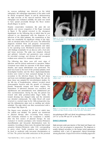
          radiograph, the radiopaque appearance of the paint could
          be clearly recognized [Figure 1a and b], demonstrating
          the  high  viscosity  of the  injected  material.  When  the
          radiograph was evaluated carefully,  the  paint was noted
          to have spread proximally  through the flexor tendon
          sheath [Figure 1a and b].

          Despite conservative treatment, the pain did not
          subside, and venous congestion of the finger increased
          in  Figure  2. The patient returned to the emergency
          department on the following day at which time he was
          referred  to  our  department  with  the  diagnosis  of  an   a            b
          infection of  the distal  phalanx.  His  examination  at that   Figure  1:  (a) Lateral  radiographic view;  (b) anteroposterior radiographic
          time was remarkable for significant edema of the digit,   view of the patient, the radiopaque appearance of the paint could be
          with an elevated white blood cell count. The differential   recognized clearly
          diagnosis  included  felon  and  pyogenic  tenosynovitis,
          and the patient was admitted immediately and taken
          to  the  operating  room.  Mid‑lateral  incisions  were  made
          with findings remarkable for an abscess with a foul odor
          and tissue necrosis. The pulp was irrigated, dressed
          and splinted. Cefazolin and gentamicin were ordered
          for microbial coverage, and dextran 40 and enoxaparin
          sodium were ordered for circulatory support.
          The  following day,  there  were  still  overt  signs  of
          infection, and the patient underwent re‑operation. Palmar
          Z‑incisions were made for exposure of the flexor tendon
          sheaths, and repeat debridement was performed. The
          paint was found at the level of the proximal phalanx. Both
          neurodigital  bundles and the  flexor digitorumprofundus
          tendon were noted to have sustained damage by lysis
          secondary  to  the  infection  [Figure  3a]. The  soft  tissue   Figure  2:  Necrosis  of the  pulp at  the  time  of  referral  to  the  plastic
          and bone were debrided aggressively [Figure 3b], and the   surgery clinic
          deep tissue  was sampled for microbiological studies. On
          the  4th  day  of  hospitalization,  cultures  revealed  mixed
          Gram‑negative bacteria with the growth of  Citrobacter
          freundii,  Morganella morgani and  Proteus vulgaris. The
          Department  of Infectious  Diseases  was  consulted, and
          ciprofloxacin and metronidazole were administered per
          their recommendation. During his stay, the patient was
          treated with daily povidone‑iodine finger baths, the
          wound dressing was changed daily,  and the  extremity
          was elevated continuously. The white blood  cell count
          returned to a normal range by the 10th postoperative day,
          and the inflammation signs such as swelling and edema
          subsided. The pulp defect was then reconstructed with a   a                     b
          cross‑finger flap [Figure 4].
                                                              Figure 3:  (a) Preoperative view of the paint along the neurovascular
          The fingers were attached for 10  days at which time   bundles and flexor tendon sheats; (b) the view after debridement
          the  pedicle was divided. The patient was transferred
          to  physical therapy  after  recovering  from  surgery,   interphalangeal (PIP) and distal interphalangeal (DIP) joints
          and eventually returned to his occupation  2  months   (10°‑15° at the PIP and 10° at the DIP).
          after the first session of physical  therapy. He was
          evaluated 12  months postinjury with Semmes‑Weinstein   DISCUSSION
          monofilaments  and two‑point  discrimination  tests  for
          sensation. According to these tests, the patient had normal   High pressure paint gun injuries of the hand and fingers are
          sensation  (Semmes‑Weinstein:  value  2.83, corresponding   very rare, but can progress to amputation. The diagnosis is
          to green color).  Tactile sensation was considered to   usually delayed secondary to the benign initial appearance
          be good to moderate with two‑point discrimination  at   combined with a lack of appropriate clinical knowledge
          6.5 mm. The patient had mild contractures at the proximal   among physicians. Surgical intervention including aggressive

          Plast Aesthet Res || Vol 2 || Issue 6 || Nov 12, 2015                                             351

