Page 232 - Read Online
P. 232
Endoscopic vision
                                                              Endoscopic surgery  consists  in  using  an  endoscope
                                                              within a natural or artificial body cavity. Conventional
                                                              microsurgery,  which  uses  an  exoscope, does  not  allow
                                                              endoscopic  vision. The 3D camera of the DaVinci  robot
                                                                                                         ®
                                                              can be used either as an exoscope or as an endoscope in
                                                              order to practice open or endoscopic telemicrosurgery.
                                                              Augmented vision
                                                              Augmented  reality consists in representing  virtual data
                                                              on a real image.  Since the invention  of this  concept by
                                                              Thomas  Caudell in  the  early  1990s, augmented  reality
                                                              has been applied to many areas. In medicine, it naturally
                                                              found technical applications using  an optical devices
                                                              and/or a camera: laparoscopy,  arthroscopy, endoscopy,
                                                                                        [2]
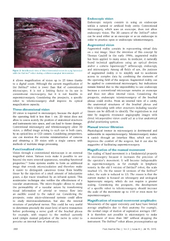
          Figure 2: Intrathoracic view of three intercostal nerves in a pig harvested   and microsurgery. Among all fields of use,  the purpose
          with the DaVinci  robot during a telemicrosurgical intervention  of augmented reality  is to simplify and to accelerate
                     ®
                                                              access to complex data by combining  the elements  of
          it  allows magnification  of vision up to 25  times  thanks   the operating field of the surgeon. Augmented reality can
          to a digital zoom. Although the current magnification  of   be applied  to conventional microsurgery, but indications
          the DaVinci  robot is lower than that of conventional   remain limited due to the impossibility to use endoscopy
                    ®
          microscopes, it is  not a limiting  factor to its  use  in   because a conventional microscope  remains an exoscope
          conventional microsurgery, but it  is  not feasible  in   and does not allow  internal vision. Considering the
          supermicrosurgery.  Considering  the  prospects,  a  specific   prospects, endoscopic telemicrosurgery  of the  brachial
          robot to telemicrosurgery shall improve its optical   plexus could evolve. From an internal view of a cavity,
          magnification capacity.                             the  anatomical structures  of the  brachial plexus and
                                                              their relationship with other structures including vascular
          Three‑dimensional vision                            tissues can be difficult to identify. The registration in real
          3D vision is required in microsurgery because the depth of   time  by magnetic  resonance angiography images  with
          the operating field is  less than 1  cm. 2D vision does not   direct intraoperative vision could act as a true anatomical
          allow to assess acutely the position of anatomical structures   global positioning system.
          and instruments into space, and can lead to tissue damage.
          Conventional microsurgery and telemicrosurgery allow 3D   Manual tremor filtration
          vision, a shifted image arriving to each eye in both cases,   Physiological tremor in microsurgery is detrimental  and
          by an optical lens or CCD camera. Considering perspectives,   unfavorable in supermicrosurgery. Telemicrosurgery makes
          one can mention the extreme miniaturization of cameras   it vanish through an interface filter, which not only
          and obtaining a 3D vision with a single camera with   improves the comfort of the surgeon, but it can also be
          methods of real‑time image processing.              suggestive of facilitating supermicrosurgery.
          Functionalized vision                               Magnification of the manual movement
          Vision through a conventional microscope is  an external   The scaling of hand movement is a fundamental property
          magnified vision. Various tools make it possible to see   in microsurgery because it increases the precision of
          beyond the mere external appearance, revealing functional   the  operator’s movement.  It  will become  indispensable
          properties.   Some  systems  enable  to form  an additional   in supermicrosurgery, as for example in lymphatic
                   [1]
          image  that reveals microcirculation and therefore make   vessels. In the old S version of DaVinci  robot, the scale
                                                                                                ®
          it  easier  to  distinguish  cancerous  tissue  from  healthy   reached 1/5. On the newer SI versions of the DaVinci
                                                                                                              ®
          tissue  by the injection of a small amount of indocyanine   robot, the scale is reduced to 1/3. The reason is that the
          green, a dye tracer visualized by an infrared system. This   current market is focused on telesurgery and urological
          noninvasive  technique  also verifies  the  effectiveness  of a   laparoscopic surgery, which  do  not  require  a greater
          vascular anastomosis.  The  micro‑Doppler allows to  study   scaling. Considering the prospects, the development
          the  permeability  of a vascular suture  by  transforming   of a specific robot to telemicrosurgery should increase
          visual information of arterial or venous flow  into   the scale of the movement up to 1/10 or even more for
          an audible  sound to the  naked ear.  Considering  the   supermicrosurgery.
          prospects, some  micro‑ultrasound systems  could allow
          to study microvascularization, but also the  internal   Magnification of manual movement amplitudes
          structure  of peripheral nerves. This  could be  very  useful   Movements of the upper extremity and hand have limited
          to determine precisely the exact level of nerve transaction   average amplitudes due to their anatomy. For example,
          before  performing  a nerve  graft  of the  brachial plexus   the normal range of motion of supination averages 180°.
          for example,  with  respect  to  the  method  currently   It is therefore not possible in microsurgery to make
          used  (simple manual palpation of the nerve in order to   a movement of more than 180°  without dropping the
          perceive an internal loss of substance).            instrument. The DaVinci  robot allows pronosupination up
                                                                                  ®
           222                                                           Plast Aesthet Res || Vol 2 || Issue 4 || Jul 15, 2015

