
Page 63 - Read Online
P. 63
Page 4 of 12 Tanner et al. Plast Aesthet Res 2023;10:11 https://dx.doi.org/10.20517/2347-9264.2022.95
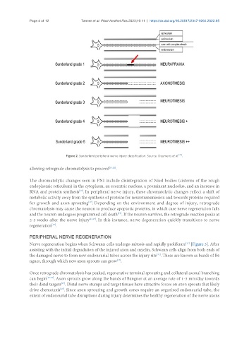
Figure 2. Sunderland peripheral nerve injury classification. Source: Deumens et al. [17] .
allowing retrograde chromatolysis to proceed [13,22] .
The chromatolytic changes seen in PNI include disintegration of Nissl bodies (cisterns of the rough
endoplasmic reticulum) in the cytoplasm, an eccentric nucleus, a prominent nucleolus, and an increase in
RNA and protein synthesis . In peripheral nerve injury, these chromatolytic changes reflect a shift of
[17]
metabolic activity away from the synthesis of proteins for neurotransmission and towards proteins required
for growth and axon sprouting . Depending on the environment and degree of injury, retrograde
[17]
chromatolysis may cause the neuron to produce apoptotic proteins, in which case nerve regeneration fails
[17]
and the neuron undergoes programmed cell death . If the neuron survives, the retrograde reaction peaks at
2-3 weeks after the nerve injury [13,17] . In this instance, nerve degeneration quickly transitions to nerve
[17]
regeneration .
PERIPHERAL NERVE REGENERATION
Nerve regeneration begins when Schwann cells undergo mitosis and rapidly proliferate [Figure 3]. After
[17]
assisting with the initial degradation of the injured axon and myelin, Schwann cells align from both ends of
[17]
the damaged nerve to form new endoneurial tubes across the injury site . These are known as bands of Bü
ngner, through which new axon sprouts can grow .
[17]
Once retrograde chromatolysis has peaked, regenerative terminal sprouting and collateral axonal branching
can begin [17-19] . Axon sprouts grow along the bands of Büngner at an average rate of 1-3 mm/day towards
their distal targets . Distal nerve stumps and target tissues have attractive forces on axon sprouts that likely
[17]
drive chemotaxis . Since axon sprouting and growth cones require an organized endoneurial tube, the
[17]
extent of endoneurial tube disruptions during injury determines the healthy regeneration of the nerve axons