
Page 34 - Read Online
P. 34
Page 10 of 13 Eftekari et al. Plast Aesthet Res 2022;9:43 https://dx.doi.org/10.20517/2347-9264.2022.33
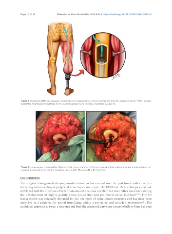
Figure 7. Illustration of the intraosseous transposition of a resected tibial nerve neuroma into the tibial medullary cavity. Photo includes
a potential intramedullary electrode for an Osseointegrated neural interface. Illustrated by Seitz AJ.
Figure 8. Intraosseous transposition following tibial nerve neuroma (left) resection with tibial corticotomy and implantation of the
proximal transected nerve into the medullary cavity (right). Photo credit to Dr. Poore SO.
DISCUSSION
The surgical management of symptomatic neuromas has evolved over the past two decades due to a
deepening understanding of peripheral nerve injury and repair. The RPNI and TMR techniques were not
developed with the intention of better outcomes of neuroma resection but were rather discovered during
the development of higher-quality neuro-prosthetics and peripheral nerve interfaces [20,25] . The IO
transposition was originally designed for the treatment of symptomatic neuroma and has since been
[1]
extended as a platform for neural interfacing within a protected and secluded environment . The
traditional approach to resect a neuroma and bury the transected nerve into a muscle belly or bone has been