
Page 11 - Read Online
P. 11
Page 6 of 17 Buncke. Plast Aesthet Res 2022;9:38 https://dx.doi.org/10.20517/2347-9264.2022.08
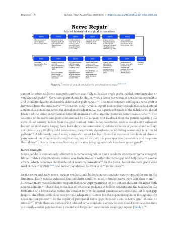
Figure 4. Timeline of surgical innovation for peripheral nerve repair [18,19,24-37] .
cannot be achieved. Nerve autografts can be successfully utilized as single grafts, cabled, interfascicular, or
[38]
vascularized grafts . Nerve autografts should be chosen from a donor nerve that is considered expendable
and would not lead to undesirable defects after graft harvest . The most common autologous nerve graft is
[38]
harvested from the sural nerve [38,42] ; however, other nerve autograft sources may include medial and lateral
antebrachial cutaneous nerve, the dorsal antebrachial nerve, the superficial branch of the radial nerve, dorsal
branch of the ulnar nerve, lateral femoral cutaneous nerve, and the posterior interosseous nerve . The
[38]
selection of the nerve autograft is determined by the surgeon with feedback from the patient regarding the
anticipated sensory deficit from the graft harvest. Sural nerve resections, such as sural nerve autograft
harvest or sural nerve biopsy, have been shown to cause sensory deficits in 92.9% of patients and sensory
symptoms (e.g., tingling cold intolerance, paresthesia, dysesthesia, or irritating sensation) in 41.1% of
[43]
patients . Additionally, sural nerve autograft harvest has been linked to increased incidents of chronic
pain, wound infection, wound complications, impact on daily life, post-operative hematoma, and deep vein
thrombosis . Due to these complications, alternative bridging materials have been investigated .
[6]
[43]
Nerve conduits
Nerve conduits were an early alternative to nerve autograft, as nerve conduits circumvent nerve autograft
harvest related complications, reduce scar tissue invasion within the nerve gap and help prevent axonal
escape, which decreases the likelihood of neuroma formation . In the 1920s, fascial and vein grafts were
[25]
used clinically by Platt [26,44] , and further popularized by Chiu et al. in the 1980s .
[45]
[29]
In the 1990s and early 2000s, various synthetic and biologic nerve conduits were proposed for use in the
[46]
literature. Early results indicated that conduits could be used to bridge nerve gaps less than 3 cm .
However, more recent literature suggests that nerve gaps measuring up to 1 cm are the limit for repair with
a nerve conduit . This is due to the lack of structural guidance in hollow conduits and the reliance on the
[47]
formation of a fibrin cable within the conduit to provide axonal guidance across the gap. At longer gap
lengths, the fibrin cable does not provide adequate structure for the regenerating axon throughout the
regeneration process . In the repair of peripheral nerve gaps beyond 1 cm, a nerve graft should be
[48]
utilized . While there are various FDA-cleared nerve conduits, a review in 2021 found that these conduits
[47]
are mostly used in gaps less than 1 cm and exhibit poorer outcomes in longer gap repairs [Table 2] .
[48]