
Page 54 - Read Online
P. 54
Page 12 of 17 Qiu et al. Plast Aesthet Res 2022;9:19 https://dx.doi.org/10.20517/2347-9264.2021.126
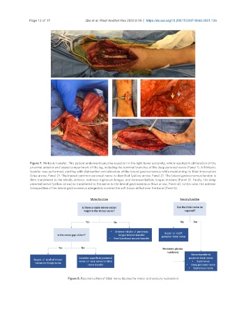
Figure 7. Ninkovic transfer. This patient underwent sarcoma resection in the right lower extremity, which resulted in obliteration of the
proximal anterior and lateral compartment of the leg, including the terminal branches of the deep peroneal nerve (Panel 1). A Ninkovic
transfer was performed, starting with disinsertion and elevation of the lateral gastrocnemius while maintaining its tibial innervation
(blue arrow, Panel 2). The injured common peroneal nerve is identified (yellow arrow, Panel 2). The lateral gastrocnemius tendon is
then transferred to the tibialis anterior, extensor digitorum longus, and extensor hallicis longus tendons (Panel 3). Finally, the deep
peroneal nerve (yellow arrow) is transferred to the nerve to the lateral gastrocnemius (blue arrow, Panel 4). In this case, the anterior
transposition of the lateral gastrocnemius adequately covered the soft tissue defect over the bone (Panel 5).
Figure 8. Reconstruction of tibial nerve injuries for motor and sensory restoration.