
Page 55 - Read Online
P. 55
Qiu et al. Plast Aesthet Res 2022;9:19 https://dx.doi.org/10.20517/2347-9264.2021.126 Page 13 of 17
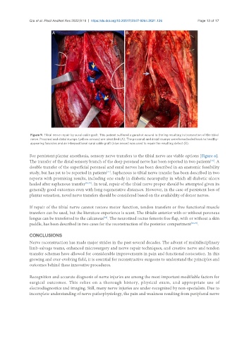
Figure 9. Tibial nerve repair by sural cable graft. This patient suffered a gunshot wound in the leg resulting in transection of the tibial
nerve. Proximal and distal stumps (yellow arrows) are identified (A). The proximal and distal stumps were breadloafed back to healthy-
appearing fascicles and an interpositional sural cable graft (blue arrow) was used to repair the resulting defect (B).
For persistent plantar anesthesia, sensory nerve transfers to the tibial nerve are viable options [Figure 8].
The transfer of the distal sensory branch of the deep peroneal nerve has been reported in two patients . A
[70]
double transfer of the superficial peroneal and sural nerves has been described in an anatomic feasibility
study, but has yet to be reported in patients . Saphenous to tibial nerve transfer has been described in two
[71]
reports with promising results, including one study in diabetic neuropathy in which all diabetic ulcers
healed after saphenous transfer [72,73] . In total, repair of the tibial nerve proper should be attempted given its
generally good outcomes even with long regenerative distances. However, in the case of persistent loss of
plantar sensation, novel nerve transfers should be considered based on the availability of donor nerves.
If repair of the tibial nerve cannot restore motor function, tendon transfers or free functional muscle
transfers can be used, but the literature experience is scant. The tibialis anterior with or without peroneus
longus can be transferred to the calcaneus . The neurotized rectus femoris free flap, with or without a skin
[74]
paddle, has been described in two cases for the reconstruction of the posterior compartment [66,75] .
CONCLUSIONS
Nerve reconstruction has made major strides in the past several decades. The advent of multidisciplinary
limb-salvage teams, enhanced microsurgery and nerve repair techniques, and creative nerve and tendon
transfer schemas have allowed for considerable improvements in pain and functional restoration. In this
growing and ever-evolving field, it is essential for reconstructive surgeons to understand the principles and
outcomes behind these innovative procedures.
Recognition and accurate diagnosis of nerve injuries are among the most important modifiable factors for
surgical outcomes. This relies on a thorough history, physical exam, and appropriate use of
electrodiagnostics and imaging. Still, many nerve injuries are under-recognized by non-specialists. Due to
incomplete understanding of nerve pathophysiology, the pain and weakness resulting from peripheral nerve