
Page 49 - Read Online
P. 49
Qiu et al. Plast Aesthet Res 2022;9:19 https://dx.doi.org/10.20517/2347-9264.2021.126 Page 7 of 17
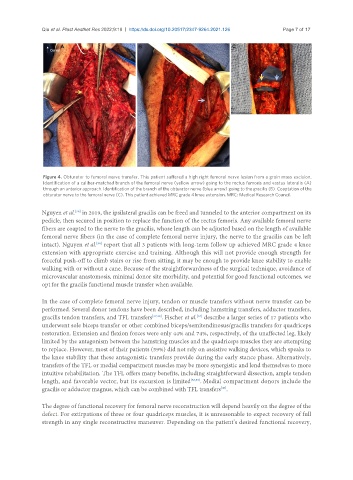
Figure 4. Obturator to femoral nerve transfer. This patient suffered a high right femoral nerve lesion from a groin mass excision.
Identification of a caliber-matched branch of the femoral nerve (yellow arrow) going to the rectus femoris and vastus lateralis (A)
through an anterior approach. Identification of the branch of the obturator nerve (blue arrow) going to the gracilis (B). Coaptation of the
obturator nerve to the femoral nerve (C). This patient achieved MRC grade 4 knee extension. MRC: Medical Research Council.
Nguyen et al. in 2019, the ipsilateral gracilis can be freed and tunneled to the anterior compartment on its
[36]
pedicle, then secured in position to replace the function of the rectus femoris. Any available femoral nerve
fibers are coapted to the nerve to the gracilis, whose length can be adjusted based on the length of available
femoral nerve fibers (in the case of complete femoral nerve injury, the nerve to the gracilis can be left
intact). Nguyen et al. report that all 3 patients with long-term follow up achieved MRC grade 4 knee
[36]
extension with appropriate exercise and training. Although this will not provide enough strength for
forceful push-off to climb stairs or rise from sitting, it may be enough to provide knee stability to enable
walking with or without a cane. Because of the straightforwardness of the surgical technique, avoidance of
microvascular anastomosis, minimal donor site morbidity, and potential for good functional outcomes, we
opt for the gracilis functional muscle transfer when available.
In the case of complete femoral nerve injury, tendon or muscle transfers without nerve transfer can be
performed. Several donor tendons have been described, including hamstring transfers, adductor transfers,
gracilis tendon transfers, and TFL transfers [37,38] . Fischer et al. describe a larger series of 17 patients who
[37]
underwent sole biceps transfer or other combined biceps/semitendinosus/gracilis transfers for quadriceps
restoration. Extension and flexion forces were only 44% and 74%, respectively, of the unaffected leg, likely
limited by the antagonism between the hamstring muscles and the quadriceps muscles they are attempting
to replace. However, most of their patients (59%) did not rely on assistive walking devices, which speaks to
the knee stability that these antagonistic transfers provide during the early stance phase. Alternatively,
transfers of the TFL or medial compartment muscles may be more synergistic and lend themselves to more
intuitive rehabilitation. The TFL offers many benefits, including straightforward dissection, ample tendon
length, and favorable vector, but its excursion is limited [38,39] . Medial compartment donors include the
[40]
gracilis or adductor magnus, which can be combined with TFL transfers .
The degree of functional recovery for femoral nerve reconstruction will depend heavily on the degree of the
defect. For extirpations of three or four quadriceps muscles, it is unreasonable to expect recovery of full
strength in any single reconstructive maneuver. Depending on the patient’s desired functional recovery,