
Page 60 - Read Online
P. 60
Page 8 of 10 Somenek. Plast Aesthet Res 2022;9:16 https://dx.doi.org/10.20517/2347-9264.2021.84
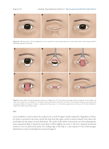
Figure 4. Cutler beard flap. (A) Flap design to the lower eyelid; (B) composite flap advanced under inferior intact eyelid margin; and (C)
following separation of the flap.
Figure 5. Hughes flap. (A) Appropriate size defect for a Hughes flap. (B) Conjunctiva and tarsus incised. Horizontal border of flap is at
least 4 mm superior to lid margin. (C) Composite flap advanced into the lower lid defect. (D) Anterior lamella reconstructed with
advancement flap. (E) Superior border or advancement flap suture 1 to 2 mm above upper border of tarsus. (F) Pedicle divided 4-6
weeks after initial reconstructive stage.
skin.
Local anesthetic is used to inject the recipient site as well the upper eyelid conjunctiva. Regardless of where
the defect is present to the lower eyelid, the flap from the upper eyelid is raised centrally from where the
tarsal plate has the largest vertical dimension. The width of the defect is measured, and when planning the
tarsoconjunctival flap, it should be noted that it will be slightly narrower to allow for adequate horizontal
tension in the reconstructed eyelid. The leading edge of the flap is 4 mm superior to the eyelid margin,
which leaves a strip of tarsal plate for structural support.