
Page 56 - Read Online
P. 56
Page 4 of 10 Somenek. Plast Aesthet Res 2022;9:16 https://dx.doi.org/10.20517/2347-9264.2021.84
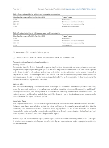
Table 1. Treatment algorithm for full-thickness lower eyelid reconstruction
Size of eyelid margin defect (% of eyelid width) Type of repair
< 25% Direct closure
25%-50% Direct closure with lateral cantholysis
50%-75% Tenzel semicircular advancement flap
> 75% Hughes tarsoconjunctival flap
Table 2. Treatment algorithm for full-thickness upper eyelid reconstruction
Size of eyelid margin defect (% of eyelid width) Type of repair
< 25% Direct closure
25%-50% Direct closure with lateral cantholysis
50%-75% Tenzel semicircular advancement flap
> 75% Cutler-beard flap
(6) Assessment of the lacrimal drainage system.
(7) To avoid corneal irritation, sutures should have knots on the cutaneous side.
Reconstruction of anterior lamellar defects
Primary closure
For anterior lamellar defects that solely require a simple ellipse for complete excision, primary closure can
be performed, especially in the upper eyelid as this area frequently has redundant skin. The tissue adjacent
to the defect should be released and undermined so that there is minimal tension on the wound closure. It is
important to orient the closure parallel to the relaxed skin tension lines (RSTLs) while the ellipses in the
periocular region should be oriented perpendicular to the RSTLs as this minimizes vertical tension and the
risk of a cicatricial ectropion or lid retraction.
Laissez faire
The concept of healing by secondary intention is usually not a reasonable option for eyelid reconstruction,
[2]
given the increased incidence of complications, including cicatricial ectropion. However, Fox and Beard
initially described this, and it has proven to be effective for relatively small medical canthal defects . This
[3]
region is concave and therefore tends to heal well if the defect is 1 cm or less in diameter and more centrally
located between the upper and lower eyelids.
Local skin flaps
[4]
Skin flaps are the favored choice over skin grafts to repair anterior lamellar defects for several reasons .
Adjacent skin has a much better match for color and texture than grafts from distant sites like the
commonly used retroauricular area. The robust blood supply allows the use of bare bone and can support
free grafts for posterior lamellar reconstruction. Additionally, there is less contraction during healing which
better supports the overall function of the periocular region.
Various flaps can be used in this region, orienting the vectors of maximal tension parallel to the lid margin.
A rotation advancement cheek flap and lateral orbital flap are reasonable and useful strategies in addition to
the options below.