
Page 59 - Read Online
P. 59
Somenek. Plast Aesthet Res 2022;9:16 https://dx.doi.org/10.20517/2347-9264.2021.84 Page 7 of 10
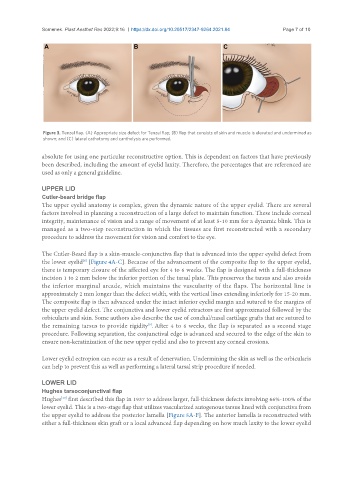
Figure 3. Tenzel flap. (A) Appropriate size defect for Tenzel flap; (B) flap that consists of skin and muscle is elevated and undermined as
shown; and (C) lateral cathotomy and cantholysis are performed.
absolute for using one particular reconstructive option. This is dependent on factors that have previously
been described, including the amount of eyelid laxity. Therefore, the percentages that are referenced are
used as only a general guideline.
UPPER LID
Cutler-beard bridge flap
The upper eyelid anatomy is complex, given the dynamic nature of the upper eyelid. There are several
factors involved in planning a reconstruction of a large defect to maintain function. These include corneal
integrity, maintenance of vision and a range of movement of at least 5-10 mm for a dynamic blink. This is
managed as a two-step reconstruction in which the tissues are first reconstructed with a secondary
procedure to address the movement for vision and comfort to the eye.
The Cutler-Beard flap is a skin-muscle-conjunctiva flap that is advanced into the upper eyelid defect from
the lower eyelid [Figure 4A-C]. Because of the advancement of the composite flap to the upper eyelid,
[8]
there is temporary closure of the affected eye for 4 to 6 weeks. The flap is designed with a full-thickness
incision 1 to 2 mm below the inferior portion of the tarsal plate. This preserves the tarsus and also avoids
the inferior marginal arcade, which maintains the vascularity of the flaps. The horizontal line is
approximately 2 mm longer than the defect width, with the vertical lines extending inferiorly for 15-20 mm.
The composite flap is then advanced under the intact inferior eyelid margin and sutured to the margins of
the upper eyelid defect. The conjunctiva and lower eyelid retractors are first approximated followed by the
orbicularis and skin. Some authors also describe the use of conchal/nasal cartilage grafts that are sutured to
[9]
the remaining tarsus to provide rigidity . After 4 to 6 weeks, the flap is separated as a second stage
procedure. Following separation, the conjunctival edge is advanced and secured to the edge of the skin to
ensure non-keratinization of the new upper eyelid and also to prevent any corneal erosions.
Lower eyelid ectropion can occur as a result of denervation. Undermining the skin as well as the orbicularis
can help to prevent this as well as performing a lateral tarsal strip procedure if needed.
LOWER LID
Hughes tarsoconjunctival flap
Hughes first described this flap in 1937 to address larger, full-thickness defects involving 66%-100% of the
[10]
lower eyelid. This is a two-stage flap that utilizes vascularized autogenous tarsus lined with conjunctiva from
the upper eyelid to address the posterior lamella [Figure 5A-F]. The anterior lamella is reconstructed with
either a full-thickness skin graft or a local advanced flap depending on how much laxity to the lower eyelid