
Page 18 - Read Online
P. 18
the role of the nerve‑muscle tissue interface in normal against external mechanical insults. Outcomes are less
nerve function. predictable than in type I lesions. Pain at rest is common
and is exacerbated by external trauma. US examination
Millesi et al. vast surgical experience with peripheral
[19]
neurolysis led to the publication of a seminal paper provides useful information on the intraneural pathology.
describing a new anatomo‑surgical classification of Type II lesions, with the exception of partial lesions
perineural and intraneural scar lesions. The classification due to a laceration or the sequelae of a nerve suture,
is a useful approach to perineural and intraneural scar correspond to Sunderland’s third‑degree lesions, which
injury because it couples each subgroup of fibrotic from the pathological standpoint include painful neuroma‑
lesions to specific types of surgical neurolysis based in‑continuity with residual function, one of the most
on scar severity. However, although intraneural lesions challenging therapeutic problems. Fourth‑ and fifth‑degree
are described in excessive detail, the clinical outcomes lesions are outside the scope of this review, as they lack
do not seem to correlate with preoperative pain residual nerve function and are managed by resection and
measurement. reconstruction.
[19]
Here we describe a simplification of Millesi et al. original
classification and propose an approach that, by correlating CLINICAL SYMPTOMS AND SIGNS
the pathological findings to clinical and imaging data, has
the potential to improve surgical treatment. The revised Patients typically report pain of four types, as described
[9]
classification encompasses two injury types, extraneural by Elliot : spontaneous pain, pressure pain, movement
and intraneural/extraneural scar lesions, based on the pain, and hypersensitivity or unpleasant skin sensation
perineural tissue changes that impair nerve gliding to light touch, including hyperesthesia, hyperpathia, and
and the intraneural problems that give rise to pain and allodynia.
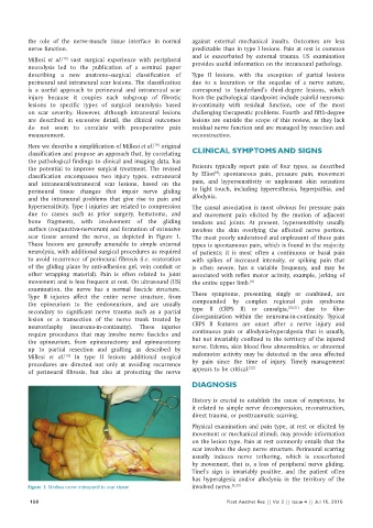
hypersensitivity. Type I injuries are related to compression The causal association is most obvious for pressure pain
due to causes such as prior surgery, hematoma, and and movement pain elicited by the motion of adjacent
bone fragments, with involvement of the gliding tendons and joints. At present, hypersensitivity usually
surface (conjunctiva‑nervorum) and formation of extensive involves the skin overlying the affected nerve portion.
scar tissue around the nerve, as depicted in Figure 1. The most poorly understood and unpleasant of these pain
These lesions are generally amenable to simple external types is spontaneous pain, which is found in the majority
neurolysis, with additional surgical procedures as required of patients; it is most often a continuous or basal pain
to avoid recurrence of perineural fibrosis (i.e. restoration with spikes of increased intensity, or spiking pain that
of the gliding plane by anti‑adhesion gel, vein conduit or is often severe, has a variable frequency, and may be
other wrapping material). Pain is often related to joint associated with reflex motor activity, example, jerking of
movement and is less frequent at rest. On ultrasound (US) the entire upper limb. [9]
examination, the nerve has a normal fascicle structure.
Type II injuries affect the entire nerve structure, from These symptoms, presenting singly or combined, are
the epineurium to the endoneurium, and are usually compounded by complex regional pain syndrome
[20,21]
secondary to significant nerve trauma such as a partial type II (CRPS II) or causalgia, due to fiber
lesion or a transection of the nerve trunk treated by disorganization within the neuroma‑in‑continuity. Typical
neurorrhaphy (neuroma‑in‑continuity). These injuries CRPS II features are onset after a nerve injury and
require procedures that may involve nerve fascicles and continuous pain or allodynia‑hyperalgesia that is usually,
the epineurium, from epineurectomy and epineurotomy but not invariably confined to the territory of the injured
up to partial resection and grafting as described by nerve. Edema, skin blood flow abnormalities, or abnormal
[19]
Millesi et al. In type II lesions additional surgical sudomotor activity may be detected in the area affected
procedures are directed not only at avoiding recurrence by pain since the time of injury. Timely management
[22]
of perineural fibrosis, but also at protecting the nerve appears to be critical.
DIAGNOSIS
History is crucial to establish the cause of symptoms, be
it related to simple nerve decompression, reconstruction,
direct trauma, or posttraumatic scarring.
Physical examination and pain type, at rest or elicited by
movement or mechanical stimuli, may provide information
on the lesion type. Pain at rest commonly entails that the
scar involves the deep nerve structure. Perineural scarring
usually induces nerve tethering, which is exacerbated
by movement, that is, a loss of peripheral nerve gliding.
Tinel’s sign is invariably positive, and the patient often
has hyperalgesia and/or allodynia in the territory of the
Figure 1: Median nerve entrapped in scar tissue involved nerve. [9,23]
158 Plast Aesthet Res || Vol 2 || Issue 4 || Jul 15, 2015