
Page 19 - Read Online
P. 19
As regards diagnostic imaging, US provides reliable in others, including patients with in‑continuity neuromas
information on the actual extent of the nerve injury (due and end‑neuromas.
for instance to a previous procedure), the amount of
scarring, and the state of the outer and inner connective SURGICAL OPTIONS
tissue layers of the nerve trunk. It thus provides an
indication for surgery by demonstrating, before the Surgical exploration, neurolysis under magnification, and
operation, the various degrees of scarring described by procedures aimed at preventing new scar formation such
Millesi et al. [19] as flap coverage and application of anti‑adhesion devices
Moreover, according to a paper of the European Society must be preceded by appropriate medical treatment and
of Musculoskeletal Radiology, musculoskeletal US seems pharmacological and physical therapy with dedicated
to be the imaging technique of choice for peripheral operators for at least six months. Although there is no
[30]
nerve structure evaluation. [24] consensus on surgery timing, surgery is generally
indicated when medical and physical therapy have failed
Most studies use US to investigate the intraneural to bring benefit.
structures and changes due to chronic compression or
trauma. In these patients, US has proven to be even Some authors have achieved pain reduction in a large
[25]
more effective than electrophysiological tests in depicting number of patients using pulsed radiofrequency before
[31]
[25]
intraneural distress. Some studies compare US findings, surgery or following a recurrence.
including signs of edema, loss of echogenicity, and Surgical treatment of these conditions begins with
fascicular echostructure before and after tunnel syndrome neurolysis. External neurolysis is performed in cases with
surgery. [26] external compression, to free the nerve from the extrinsic
compression. This may involve either accessing only the
Padua et al. group has advanced an interesting proposal
[27]
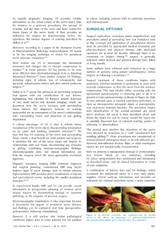
that agrees with our classification of scar lesions, epineurium (epineurotomy) or removing part or all of it
highlighting that valuable US features include depiction (partial or total epineurectomy) as shown in Figure 2a. Only
of very small nerves and dynamic imaging, which can in very selected cases is internal neurolysis performed, to
treat an intraoperative iatrogenic injury or postoperative
document how the nerve interacts with surrounding scar recurrence between fascicles. The procedure begins
tissue. Indeed, key diagnostic features of scarring with identification of the normal proximal and distal
neuropathy are an assessment of the nerve’s relationships nerve portions; the nerve is then mobilized above and
with surrounding tissue and depiction of any gliding
impairment. below the injury site and its course toward the injury site
is carefully dissected free of external scarring, points of
A critical advantage of US is that it affords direct tethering, or abnormalities.
visualization of the nerve injury, thus providing information The second step involves the relocation of the nerve
on its cause and enabling treatment selection. We tract involved by neurolysis to a “soft” vascularized bed
[27]
thus feel that US scanning of the nerve and surrounding [30]
tissue entails a dual benefit for both patient and surgeon: enabling gliding. Other procedures use vascularized or
it identifies the site of the nerve injury and depicts its nonvascularized autologous tissue or an anti‑adhesion gel.
However, anti‑adhesion devices, flaps, or other autologous
relationships with scar tissue, documenting any obstacles tissues are not unequivocally recommended.
to gliding. Combining anatomo‑sonographic findings,
electromyography data, and clinical information can Here we propose a management strategy of posttraumatic
help the surgeon select the most appropriate treatment scar lesions based on two mainstays, including
approach. (1) lesion categorization into extraneural and intraneural
as described above, and (2) clinical information in terms
Magnetic resonance imaging (MRI) enhances diagnosis of pain symptoms.
and surgical planning; conventional MRI may depict
indirect signs of nerve damage such as edema whereas A combination of history data and US findings, which
high‑resolution MRI provides direct visualization of injured document the intraneural injury in a very early phase,
and scar‑tethered nerves, including the smaller peripheral supplies critical work‑up information and provides an
branches. [28,29] indication for external neurolysis versus a more extensive
In experienced hands, MRI and US can provide crucial
information in preoperative planning of revision nerve
release surgery by documenting residual or recurrent
pathology or the sequelae of previous surgery.
Electromyography examination is also important because
it documents the degree of peripheral nerve distress,
and findings can be compared over time (preoperative,
postoperative, follow‑up examination). a b
However, it is still unclear why similar pathological Figure 2: (a) External neurolysis and epineurectomy on median
nerve at the elbow; (b) application of carboxy‑methylcellulose/
conditions induce pain in some patients but are painless phosphatidylethanolamine gel on median nerve after neurolysis
Plast Aesthet Res || Vol 2 || Issue 4 || Jul 15, 2015 159