
Page 13 - Read Online
P. 13
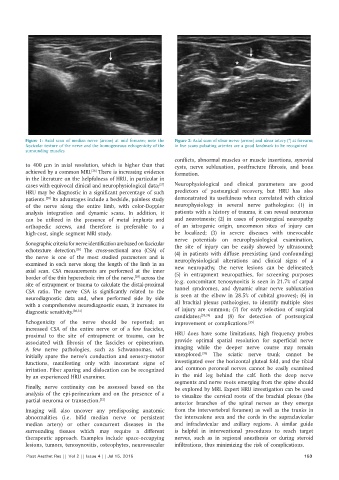
Figure  1: Axial scan of median nerve  (arrow) at mid  forearm; note the   Figure 2: Axial scan of ulnar nerve (arrow) and ulnar artery (*) at forearm;
          fascicular texture of the nerve and the homogeneous echogenicity of the   in live scans pulsating arteries are a good landmark to be recognized
          surrounding muscles
                                                              conflicts, abnormal muscles or muscle insertions, synovial
          to 400  μm in axial resolution, which is higher than that   cysts, nerve subluxation, postfracture fibrosis, and bone
          achieved by a common MRI.  There is increasing evidence   formation.
                                 [26]
          in the literature on the helpfulness of HRU, in particular in
          cases with equivocal clinical and neurophysiological data;    Neurophysiological and clinical parameters are good
                                                         [27]
          HRU may be diagnostic in a significant percentage of such   predictors of  postsurgical  recovery,  but  HRU  has  also
          patients.  Its advantages include a bedside, painless study   demonstrated its usefulness when correlated with clinical
                 [28]
          of the nerve along the entire limb, with color‑Doppler   neurophysiology  in  several  nerve  pathologies: (1) in
          analysis integration and dynamic scans. In addition, it   patients with a history of trauma, it can reveal neuromas
          can  be  utilized  in  the  presence  of  metal  implants  and   and neurotmesis;  (2) in  cases of postsurgical neuropathy
          orthopedic  screws,  and  therefore  is  preferable  to  a   of an iatrogenic origin,  uncommon  sites  of injury can
          high‑cost, single segment MRI study.                be  localized;  (3) in  severe  diseases  with  unevocable
                                                              nerve potentials on neurophysiological examination,
          Sonographic criteria for nerve identification are based on fascicular   the site of injury can be easily showed by ultrasound;
          echotexture detection.   The  cross‑sectional area (CSA) of   (4) in  patients  with  diffuse  preexisting  (and confounding)
                            [26]
          the nerve is one of the most studied parameters and is   neurophysiological alterations  and clinical signs  of a
          examined in each nerve along the length of the limb in an   new  neuropathy,  the  nerve  lesions  can be  delineated;
          axial scan. CSA measurements are performed at the inner
          border of the thin hyperechoic rim of the nerve,  across the   (5)  in entrapment neuropathies, for screening  purposes
                                                [29]
          site of entrapment or trauma to calculate the distal‑proximal   (e.g. concomitant tenosynovitis is seen in 21.7% of carpal
          CSA  ratio.  The  nerve  CSA  is  significantly  related  to  the   tunnel  syndromes,  and dynamic  ulnar nerve  subluxation
          neurodiagnostic data and, when performed side by side   is  seen  at  the  elbow in  28.5%  of cubital grooves);  (6) in
          with a comprehensive neurodiagnostic exam, it increases its   all brachial  plexus  pathologies,  to  identify  multiple  sites
          diagnostic sensitivity. [30,31]                     of injury  are  common;  (7) for early  selection  of surgical
                                                              candidates; [33,34]  and  (8)  for detection of postsurgical
          Echogenicity  of the  nerve  should be  reported; an   improvement or complications. [35]
          increased  CSA  of  the  entire  nerve  or  of  a  few  fascicles,
          proximal  to the site  of entrapment  or trauma,  can be   HRU does have some limitations, high frequency probes
          associated with fibrosis of the fascicles or epineurium.   provide optimal spatial resolution for superficial nerve
          A  few  nerve  pathologies,  such as  Schwannomas,  will   imaging while the deeper nerve course may remain
                                                                        [36]
          initially  spare  the  nerve’s  conduction and sensory‑motor   unexplored.   The  sciatic  nerve  trunk  cannot  be
          functions, manifesting  only with inconstant signs of   investigated over the horizontal gluteal fold, and the tibial
          irritation. Fiber sparing and dislocation can be recognized   and common peroneal nerves cannot be easily examined
          by an experienced HRU examiner.                     in the mid leg behind the calf. Both the deep nerve
                                                              segments and nerve roots emerging from the spine should
          Finally,  nerve continuity  can be  assessed  based on the   be explored by MRI. Expert HRU investigation can be used
          analysis of the epi‑perineurium and on the presence of a   to visualize the cervical roots of the brachial plexus (the
          partial neuroma or transection. [32]                anterior branches of the spinal nerves as they emerge
          Imaging  will  also  uncover any  predisposing  anatomic   from  the  intervertebral  foramen)  as  well  as  the  trunks  in
          abnormalities  (i.e.  bifid median nerve or persistent   the interscalene area and the cords in the supraclavicular
          median  artery) or other  concurrent  diseases  in  the   and  infraclavicular  and  axillary  regions.  A  similar  guide
          surrounding  tissues  which  may  require  a  different   is helpful in interventional procedures to reach target
          therapeutic approach. Examples include space‑occupying   nerves,  such  as  in  regional  anesthesia  or  during  steroid
          lesions, tumors, tenosynovitis, osteophytes, neurovascular   infiltrations, thus minimizing the risk of complications.
          Plast Aesthet Res || Vol 2 || Issue 4 || Jul 15, 2015                                             153