
Page 10 - Read Online
P. 10
the indications and correct timing for each instrumental corresponds to neuropraxia; (2) stage II corresponds
examination will be reviewed, with a specific focus on to axonotmesis; and (3) stages III, IV, and V correspond
innovative methods and future prospects. to neurotmesis [Table 1], with impairment of the
endoneurium, perineurium, and epineurium.
CLASSIFICATION OF PERIPHERAL The distinction between the different types of injury
NERVE INJURIES is not always precise. Clinical evaluation benefits from
instrumental approaches to discriminate severity at an
The most commonly used classification for peripheral earlier stage, thus allowing for appropriate and timely
nerve injuries is that by Seddon, and Sunderland. The treatment.
[3]
[4]
Seddon classification places injuries into three basic types:
neurapraxia, axonotmesis, and neurotmesis. CLINICAL APPROACH
Neurapraxia (praxis: to do, to perform): the nerve axons
are intact but cannot transmit impulses. This occurs Patient age, mechanism of injury and associated vascular
secondary to ischemic damage with temporary myelin and soft tissue injuries strongly influence the extent
sheath damage. Without myelin, there is an alteration of recovery of the injured nerve. These elements are of
of “saltatory conduction” across the nodes of Ranvier great importance and are the primary details collected
with subsequent slowed or blocked nerve conduction. in the clinical history. A detailed examination includes
Neuropraxia is the mildest form of nerve injury; “Saturday evaluation of pain and muscular strength and sensory
night” radial palsy and entrapment neuropathies like testing in the territory of the injured nerve. The
carpal tunnel syndrome is good example for this homologous contralateral and other ipsilateral preserved
condition. [5,6] Nerve recovery occurs after remyelination nerves are used for comparison, particularly in polytrauma
and sensory‑motor functions can usually completely patients. Appropriate motor and sensory evaluation
[11]
restored within days to weeks. [7] is mandatory to identify injuries to sensitive, motor,
Axonotmesis (tmesis: to cut): the axons are damaged and mixed nerves; early and late signs of autonomic
or destroyed, but most of the connective scaffold disorders should also be evaluated, including vasomotor
(endoneurium, perineurium, and epineurium) remains disorders and trophic alteration of the skin, nails, and
intact. Axonotmesis is commonly seen in crush and stretch subcutaneous tissue. [11,12] Both negative (e.g. hypoesthesia,
[8]
injuries. After injury, anterograde Wallerian degeneration muscle weakness, and atrophy) and positive symptoms
of the distal axonal fibers is completed within a few days. (e.g. dysesthesia, pain, fasciculations) due to loss of
nerve function or inappropriate spontaneous activity,
Neurotmesis: the nerve trunk is disrupted and loses respectively, should be noted.
anatomical continuity. Neurotmesis represents the most
severe form of injury with disruption of the axons, myelin The simplest standardized clinical evaluation of a
sheath, and connective tissues. It may occur following sharp cutaneous somatic sensitivity is the test of the pain
injuries, massive trauma, or severe traction that partially pathway (the patient’s ability to perceive the touch of a
[13]
or completely interrupts nerve continuity. In order to sharp object). Clinicians and surgeons generally refer to
[9]
enhance the chances for reinnervation after neurotmesis, cutaneous nociception because of less lower overlap of
surgical nerve repair is mandatory. Without surgery, innervating territories when compared to tactile sensation.
[10]
uncontrolled axonal re‑growth will generate a neuroma.
Hypoesthesia generally involves all superficial and deep
The Sunderland classification includes five stages somatosensory systems (tactile, thermal, pain, and
and identifies three types of neurotmesis: (1) stage I proprioception); anatomical charts and diagrams help to
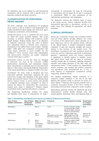
Table 1: Classification of peripheral nerve injuries according to Seddon and Sunderland
Type of injury Type of Injury Major structure Prognosis Neurodiagnostic findings Requirement
Seddon classification Sunderland involved for surgical
classification intervention
Neuropraxia I Myelin Good Slower conduction velocity None
or conduction block; EMG
with no fibrillation, reduced
recruitment and fast firing
Axonotmesis II Myelin, Fair Reduced CMAP and Depends on
Axons (depending on how many SNAP amplitudes; EMG extension of
fibers are involved) with fibrillation, reduced the lesion
recruitment and fast firing
Neurotmesis III, Myelin, Poor Reduced or absent CMAP Often requires
IV, Axons, (depending on how many and SNAP; EMG with surgical repair
V Endoneurium fibers are involved) fibrillation and motor units
Perineurium loss
Epineurium
EMG: Electromyography, CMAP: Compound muscle action potential, SNAP: Sensory nerve action potential
150 Plast Aesthet Res || Vol 2 || Issue 4 || Jul 15, 2015