
Page 80 - Read Online
P. 80
Pang et al. Plast Aesthet Res 2021;8:49 https://dx.doi.org/10.20517/2347-9264.2021.42 Page 7 of 11
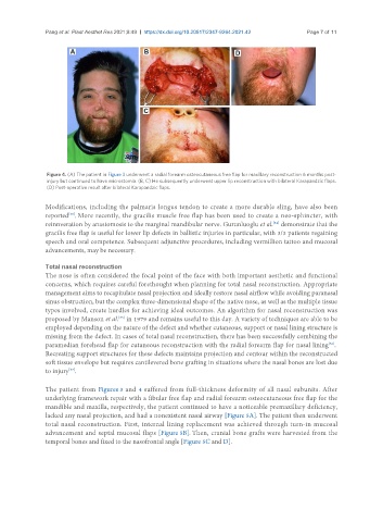
Figure 4. (A) The patient in Figure 3 underwent a radial forearm osteocutaneous free flap for maxillary reconstruction 6 months post-
injury but continued to have microstomia. (B, C) He subsequently underwent upper lip reconstruction with bilateral Karapandzic flaps.
(D) Post-operative result after bilateral Karapandzic flaps.
Modifications, including the palmaris longus tendon to create a more durable sling, have also been
[33]
reported . More recently, the gracilis muscle free flap has been used to create a neo-sphincter, with
reinnveration by anastomosis to the marginal mandibular nerve. Gurunluoglu et al. demonstrate that the
[34]
gracilis free flap is useful for lower lip defects in ballistic injuries in particular, with 3/3 patients regaining
speech and oral competence. Subsequent adjunctive procedures, including vermillion tattoo and mucosal
advancements, may be necessary.
Total nasal reconstruction
The nose is often considered the focal point of the face with both important aesthetic and functional
concerns, which requires careful forethought when planning for total nasal reconstruction. Appropriate
management aims to recapitulate nasal projection and ideally restore nasal airflow while avoiding paranasal
sinus obstruction, but the complex three-dimensional shape of the native nose, as well as the multiple tissue
types involved, create hurdles for achieving ideal outcomes. An algorithm for nasal reconstruction was
proposed by Manson et al. in 1979 and remains useful to this day. A variety of techniques are able to be
[35]
employed depending on the nature of the defect and whether cutaneous, support or nasal lining structure is
missing from the defect. In cases of total nasal reconstruction, there has been successfully combining the
paramedian forehead flap for cutaneous reconstruction with the radial forearm flap for nasal lining .
[36]
Recreating support structures for these defects maintains projection and contour within the reconstructed
soft tissue envelope but requires cantilevered bone grafting in situations where the nasal bones are lost due
[37]
to injury .
The patient from Figures 3 and 4 suffered from full-thickness deformity of all nasal subunits. After
underlying framework repair with a fibular free flap and radial forearm osteocutaneous free flap for the
mandible and maxilla, respectively, the patient continued to have a noticeable premaxillary deficiency,
lacked any nasal projection, and had a nonexistent nasal airway [Figure 5A]. The patient then underwent
total nasal reconstruction. First, internal lining replacement was achieved through turn-in mucosal
advancement and septal mucosal flaps [Figure 5B]. Then, cranial bone grafts were harvested from the
temporal bones and fixed to the nasofrontal angle [Figure 5C and D].