
Page 77 - Read Online
P. 77
Page 4 of 11 Pang et al. Plast Aesthet Res 2021;8:49 https://dx.doi.org/10.20517/2347-9264.2021.42
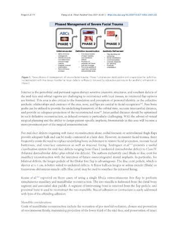
Figure 1. Three phases of management of severe facial trauma. Phase 1 emphasizes stabilization and preparation for definitive
reconstruction with free tissue transfer for major defects in Phase 2, followed by adjunctive procedures for aesthetic refinement in
Phase 3.
Injuries to the periorbital and perinasal region disrupt sensitive anatomic structures, and resultant defects of
the mid-face and orbital regions are challenging to reconstruct with local tissues, as rotational flap options
are limited. This area is also critical to the foundation and perception of personal identity, as the collective
[10]
aesthetic relationships and contours of the eyes, nose, and lips are central to facial recognition . Free bone
grafts can be utilized to provide the underlying framework of the orbital rims, recreate intercanthal distance,
and provide an adequate projection of the reconstructed nose . Intercanthal distance should be optimized
[9]
in early definitive reconstruction, as delayed revision is particularly challenging. With the advent of virtual
surgical planning and the ability to design patient-specific implants, biomaterials in this area will become a
more prominent part of the surgical armamentarium.
For mid-face defects requiring soft tissue reconstruction alone, radial forearm or anterolateral thigh flaps
provide adequate bulk and can be easily contoured at a later date. However, in massive facial trauma, there
frequently exists the need to replace underlying bony architecture to restore facial projection, recreate facial
buttresses, and resurface cutaneous as well as mucosal lining. Rodriguez et al. presents a useful
[19]
classification system for mid-face defects ranging from Class I (unilateral dentoalveolar defect) to Class IV
(bilateral dentoalveolar defect plus orbital rim defects). The authors exclusively used fibula or iliac crest for
maxillary reconstruction with the intention of future osseointegrated dental implants. In particular, for
bilateral defects, the longer pedicle of the fibular free flap is advantageous. The iliac crest pedicle, which is
shorter at 4-5 cm, is better suited to unilateral defects. A flexor hallucis longus or soleus muscle (fibula) or
transversus abdominus muscle cuffs (iliac crest) may be used to resurface the intraoral lining.
Krane et al. reported on three cases of using a single fibula osteocutaneous free flap to perform
[20]
simultaneous maxillary and mandibular reconstruction. The neo-maxilla is fashioned from the distal bony
segment and associated skin paddle. A segment of intervening bone is removed from the flap pedicle, and
proximal bone is used to reconstruct the neo-mandible. Buccal adhesion or contracture is easily addressed
with lysis of the offending adhesion.
Mandible considerations
Goals of mandibular reconstruction include the recreation of pre-morbid occlusion, closure and prevention
of orocutaneous fistula, maintaining projection of the lower third of the mid-face, and preservation of intact