
Page 78 - Read Online
P. 78
Pang et al. Plast Aesthet Res 2021;8:49 https://dx.doi.org/10.20517/2347-9264.2021.42 Page 5 of 11
nerves for optimal speech and swallow function. Occasionally, facial trauma may result in fractured but
preserved mandibular bone attached to the overlying periosteum, which is amenable to rigid fixation
without the need for additional free tissue. For example, such defects may occur in the setting of self-
[2]
inflicted submental gunshot wounds .
The fibular osteocutaneous free flap has become the workhorse of mandibular reconstruction for segmental
defects in the setting of trauma [2,21,22] . While non-vascularized bone grafts may be used for bony defects <
5 cm, it is recommended to utilize free vascularized osseous tissue for bony defects > 5 cm . Up to 25 cm of
[23]
bone can be harvested while preserving the proximal and distal 6 cm at the knee and ankle joint,
respectively. Moreover, a generous skin paddle based on a septo- or musculo-cutaneous perforator may be
[24]
harvested, and multiple skin paddles may be designed if more than one perforator is captured . Rigid
fixation to the native mandible combined with meticulous watertight closure ensures optimal results.
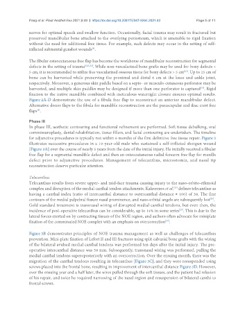
Figure 2A-D demonstrate the use of a fibula free flap to reconstruct an anterior mandibular defect.
Alternative donor flaps to the fibula for mandible reconstruction are the parascapular and iliac crest free
flaps .
[2]
Phase III
In phase III, aesthetic contouring and functional refinement are performed. Soft tissue debulking, oral
commissuroplasty, dental rehabilitation, tissue fillers, and facial contouring are undertaken. The timeline
for adjunctive procedures is typically not within 6 months of the first definitive free tissue repair. Figure 3
illustrates successive procedures in a 19-year-old male who sustained a self-inflicted shotgun wound
[Figure 3A] over the course of nearly 5 years from the date of the initial injury. He initially received a fibular
free flap for a segmental mandible defect and then an osteocutaneous radial forearm free flap for maxilla
defect prior to adjunctive procedures. Management of telecanthus, microstomia, and nasal tip
reconstruction deserve particular attention.
Telecanthus
Telecanthus results from severe upper- and mid-face trauma causing injury to the naso-orbito-ethmoid
[25]
complex and disruption of the medial canthal tendon attachments. Kalavrezos et al. defines telecanthus as
having a canthal index (ratio of intercanthal distance to outercanthal distance × 100) of 38. The fine
[26]
contours of the medial palpebral fissure nasal prominence, and naso-orbital angels are subsequently lost .
Gold standard treatment is transnasal wiring of disrupted medial canthal tendons, but even then, the
incidence of post-operative telecanthus can be considerable, up to 14% in some series . This is due to the
[25]
lateral forces exerted on by contracting tissues of the NOE region, and authors often advocate for miniplate
fixation of the comminuted NOE complex with an emphasis on overcorrection .
[27]
Figure 3B demonstrates principles of NOE trauma management as well as challenges of telecanthus
prevention. Mini-plate fixation of Lefort II and III fractures using split calvarial bone grafts with the wiring
of the bilateral avulsed medial canthal tendons was performed ten days after the initial injury. The pre-
operative intercanthal distance was 58 mm. Subsequently, transnasal wiring was performed, pulling the
medial canthal tendons superoposteriorly with an overcorrection. Over the ensuing month, there was the
migration of the canthal tendons resulting in telecanthus [Figure 3C], and they were resuspended using
screws placed into the frontal bone, resulting in improvement of intercanthal distance Figure 3D. However,
over the ensuing year and a half later, the wires pulled through the soft tissues, and the patient had relaxion
of his repair, and twice he required narrowing of the nasal region and resuspension of bilateral canthi to
frontal screws.