
Page 85 - Read Online
P. 85
Page 8 of 12 Pandey et al. Plast Aesthet Res 2021;8:47 https://dx.doi.org/10.20517/2347-9264.2021.61
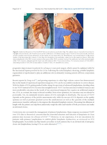
Figure 5. Anatomical dissection of the thin TDAP (thoracodorsal artery perforator) flap. The cadaver was pre-injected with red latex to
highlight arterial anatomy. (A) Possible sources of skin perforators in the thoracodorsal region. Posterior intercostal vessels, which are
another possible source of skin perforators in this region, are not shown here. (B) The perforator was located anterior to the lateral
border of the latissimus dorsi muscle. A thin flap was elevated on this perforator, superficial to the superficial fascia. While the
descending branch of the TDA is most commonly the source of the TDAP, proximal tracing of this perforator led to the serratus branch
of the TDA. However, adequate pedicle length was still achieved. TDA: Thoracodorsal artery; TB: transverse branch of TDA; DBTD:
descending branch of TDA; SAB: serratus anterior branch of TDA; LTA: lateral thoracic artery.
progressive improvement in patients for as long as 3 years post-surgery, which cannot be explained solely by
the mechanical bypass provided by the LVAs. Following the initial lymphatic shunting, possible lymphatic
regeneration is hypothesized to play an additional role in ultimately weaning patients off from compression
garments.
Recent reports by Yang et al. , and growing experience in other high-volume centers has demonstrated
[76]
that in the hands of experienced supermicrosurgeons, LVA can be successful in moderate-to-severe disease.
With the limits of LVA getting pushed further along the spectrum of advanced lymphedema, deciding when
to use VLVT instead of LVA becomes less straightforward. VLVT has less associated technical nuances and
more predictable outcomes in the hands of less experienced surgeons but requires an additional surgical
site. LVA, in contrast, has many technical variables. Even when optimized, its outcomes may not always be
predictable. Yet, the minimally invasive nature of LVA outweighs its drawbacks. The success of VLVT
depends on the patency of the pedicle anastomosis only, while the success of LVA depends on the quality
(patency as well as pressure gradient across the anastomoses) as well as the quantity of the multiple
anastomoses (number sufficient to decompress the distended lymphatic system). Discussing this dilemma at
length with the patient can help them judiciously weigh the risks and benefits of both procedures and make
an informed choice.
Controversy also surrounds the management of primary lymphedema. Many surgeons recommend VLNT
over LVA due to theoretical concerns that the abnormal structure and function of lymphatics in these
patients may decrease the efficacy of LVA [2,77,78] . However, in our experience, it is not uncommon for
patients with primary lymphedema to exhibit global lymphatic dysfunction, as evidenced on ICG
lymphography. It precludes any flap transfer procedure in such patients due to an elevated risk of iatrogenic
donor-site lymphedema, leaving LVA as a safer alternative [64,79,80] .