
Page 23 - Read Online
P. 23
Page 4 of 17 Preto et al. Plast Aesthet Res. 2025;12:28 https://dx.doi.org/10.20517/2347-9264.2025.26
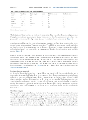
Table 1. Female sexual function index - FSFI - score interpretation
Domain Questions Score range Factor Minimum score Maximum score
Desire 1, 2 1-5 0,6 1.2 6.0
Arousal 3, 4, 5, 6 0-5 0,3 0 6.0
Lubrication 7, 8, 9, 10 0-5 0,3 0 6.0
Orgasm 11, 12, 13 0-5 0,4 0 6.0
Satisfaction 14, 15, 16 0 (or 1)-5 0,4 0.8 6.0
Pain 17, 18, 19 0-5 0,4 0 6.0
Full scale score range 2.0 36.0
FSFI: Female sexual function index.
The first phase of the procedure was the demolitive phase, involving bilateral orchiectomy and penectomy.
During this phase, tissues and anatomical structures necessary for the subsequent reconstructive phase were
preserved. A perineoscrotal skin flap was elevated, and the penile shaft skin was preserved. [Figure 1].
A urethral mucosal flap was also preserved to create the neovulvar vestibule, along with retraction of the
urethral meatus and meatoplasty. The preputial skin flap (if available), the neurovascular bundle attached to
the dorsal portion of the tunica albuginea, and the dorsocentral portion of the glans (reconfigured according
to Preecha’s technique) were preserved for the construction of the clitoral complex, clitoral hood, and labia
minora. [Figure 2].
Once the neovaginal cavity was created (between the rectal wall and the urethroprostatic plane, following
Denonvilliers’ fascia), it was lined with appropriately approximated and sutured perineoscrotal and penile
skin flaps. In cases of limited skin availability, a full-thickness skin graft harvested from excess scrotal skin
was applied to ensure maximum neovaginal depth. After lining the vaginal cavity, the neovulvar vestibule
(including the clitoral complex/labia minora, urethral meatus, and vaginal introitus) was completed, and the
labia majora were created and sutured. (Figure 3 - for more details about the surgical technique, see
Supplementary Figures 1-3)
Postoperative management
At the end of the surgical procedure, a vaginal dilator was placed inside the neovaginal cavity, and a
compressive dressing applied for five days. On postoperative day 5, the compressive dressing, vaginal stent,
and catheter were removed. Initially, a soft dilator was used at night for the first 15 days, followed by rigid
dilators, with three one-hour dilation sessions per day, for at least nine months. The first postoperative visit
was scheduled around 21 days after the procedure. The follow-up visits were scheduled at 3, 6, and 12
months after surgery to evaluate the healing process, the findings of the dilation regimen, and to rule out
complications or the need for surgical revision. Sexual activity was allowed starting from the second month
post-surgery.
Statistical analysis
The normality of variable distributions was tested by the Kolmogorov-Smirnov test. Categorical variables
were described using frequencies and percentages. For continuous variables with a normal distribution,
mean and standard deviation (SD) were reported, whereas variables with a non-normal distribution were
described using median and interquartile range (IQR). Differences between groups were assessed by the chi-
square test or Fisher’s exact test for discrete variables. For continuous variables, differences between groups
were assessed by the Student’s t-test for normally distributed and the Mann-Whitney U test or the
Wilcoxon test (respectively for independent and paired samples) for non-normally distributed variables. A