
Page 95 - Read Online
P. 95
Scaglioni et al. Plast Aesthet Res 2019;6:27 I http://dx.doi.org/10.20517/2347-9264.2019.41 Page 7 of 10
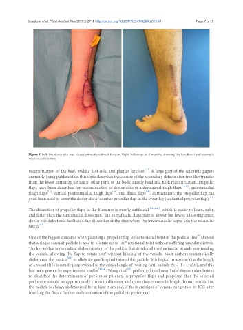
Figure 7. Left: the donor site was closed primarily without tension. Right: follow-up at 4 months, showing the functional and cosmetic
result is satisfactory
[17]
reconstruction of the heel, middle foot sole, and plantar forefoot . A large part of the scientific papers
currently being published on this topic describes the closure of the secondary defects after free flap transfer
from the lower extremity for use in other parts of the body, mostly head and neck reconstruction. Propeller
flaps have been described for reconstruction of donor sites of anterolateral thigh flaps [12,18] , anteromedial
[18]
[19]
[20]
thigh flaps , vertical posteromedial thigh flaps , and fibula flaps . Furthermore, the propeller flap has
[11]
even been used to cover the donor site of another propeller flap in the lower leg (sequential propeller flap) .
The dissection of propeller flaps in the literature is mostly subfascial [7,8,14,21] , which is easier to learn, safer,
and faster than the suprafascial dissection. The suprafascial dissection is slower but leaves a less important
donor-site defect and facilitates flap dissection at the sites where the intermuscular septa join the muscular
[22]
fascia .
[8]
One of the biggest concerns when planning a propeller flap is the torsional twist of the pedicle. Teo showed
that a single vascular pedicle is able to tolerate up to 180° rotational twist without suffering vascular distress.
The key to that is the radical skeletonization of the pedicle that divides all the fine fascial strands surrounding
the vessels, allowing the flap to rotate 180° without kinking of the vessels. Most authors systematically
[8,9]
skeletonize the pedicle to allow for gentle spiral twist of the pedicle. It is logical to assume that the length
of a vessel (l) is inversely proportional to the critical angle of twisting (Δt), namely Δt = [l × (1/Δt)], and this
[25]
has been proven by experimental studies [23,24] . Wong et al. performed nonlinear finite element simulations
to elucidate the determinants of perforator patency in propeller flaps and proposed that the selected
perforator should be approximately 1 mm in diameter and more than 30 mm in length. In our institution,
the pedicle is always skeletonized for at least 3 cm and, if there are signs of venous congestion in ICG after
insetting the flap, a further skeletonization of the pedicle is performed.