
Page 91 - Read Online
P. 91
Scaglioni et al. Plast Aesthet Res 2019;6:27 I http://dx.doi.org/10.20517/2347-9264.2019.41 Page 3 of 10
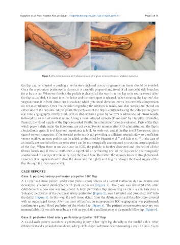
Figure 1. Wound dehiscence with plate exposure after plate osteosynthesis of a lateral malleolus
the flap can be adjusted accordingly. Perforators enclosed in scar or granulation tissue should be avoided.
Once the appropriate perforator is chosen, it is carefully prepared and freed of all muscular side branches
for at least 2 cm. Wherever feasible, the pedicle is cleaned all the way from the flap to its source vessel. After
the flap is islanded, it is inset into the defect and the tourniquet is released. When rotating the flap 180°, the
surgeon turns it in both directions to evaluate which rotational direction exerts less extrinsic compression
on venae comitantes. Once the decision regarding the rotation is made, two skin sutures are placed on
either side of the flap axis. At this point, the perfusion of the flap is controlled using the indocyanine green
real-time angiography. Firstly, 2 mL of ICG (Indocyanine green by Verdy®) is administered intravenously,
followed by 10 mL of normal saline. Using a near-infrared camera (Fluobeam® by Fluoptics Grenoble,
France), the blood supply of the flap is recorded. Firstly, the arterial perfusion is evaluated. Parts of the flap,
which present dark under the Fluobeam, are cut away. Twenty minutes after ICG administration, the flap is
checked once again. It is of foremost importance to look for wash-out, and, if the flap is still fluorescent, it is a
sign of venous congestion. If the isolated perforator is not providing a sufficient arterial inflow or a sufficient
[12]
[9]
venous outflow, an extra pedicle can be added, as described by Pignatti et al. and Iida et al. In the case of
an insufficient arterial inflow, an extra artery can be microsurgically anastomosed to a second arterial pedicle
of the flap. When there is no wash-out on ICG, the pedicle is further dissected and cleaned of all the
fibrous bands and, if this is insufficient, a superficial or perforating vein of the flap can be microsurgically
anastomosed to a recipient vein to increase the blood flow. Thereafter, the wound closure is straightforward.
However, it is important not to close the donor site too tightly as it might endanger the blood supply of the
flap through the tourniquet effect.
CASE REPORTS
Case 1: peroneal artery perforator propeller 180° flap
A 41-year-old male patient underwent plate osteosynthesis of a lateral malleolus due to trauma and
developed a wound dehiscence with plate exposure [Figure 1]. The plate was removed and, after
debridement, a new one was implanted. A local perforator flap measuring 16 cm × 4 cm, based on a
Y-shaped perforator of the peroneal artery perforator [Figure 2], was harvested and propelled 180° into
the defect [Figure 3]. In this way, the soft tissue defect from the debridement and the plate were covered
with an undamaged tissue. After the inset of the flap, an intraoperative ICG angiography was performed,
confirming a good blood perfusion of the whole flap [Figure 4]. The patient’s postoperative recovery was
unremarkable. He was able to ambulate with no restriction and limitation at six-month follow-up [Figure 5].
Case 2: posterior tibial artery perforator propeller 180° flap
A 24-old-male patient sustained a penetrating injury of her right leg, dorsally to the medial ankle. After
debridement and a period of wound care, a deep, circle-shaped soft tissue defect measuring 4 cm × 3.5 cm × 2.5 cm