
Page 92 - Read Online
P. 92
Page 4 of 10 Scaglioni et al. Plast Aesthet Res 2019;6:27 I http://dx.doi.org/10.20517/2347-9264.2019.41
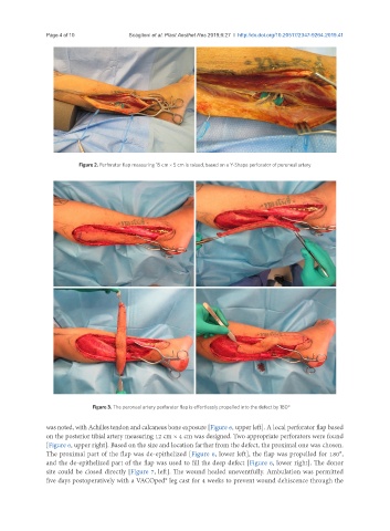
Figure 2. Perforator flap measuring 15 cm × 5 cm is raised, based on a Y-Shape perforator of peroneal artery
Figure 3. The peroneal artery perforator flap is effortlessly propelled into the defect by 180°
was noted, with Achilles tendon and calcaneus bone exposure [Figure 6, upper left]. A local perforator flap based
on the posterior tibial artery measuring 12 cm × 4 cm was designed. Two appropriate perforators were found
[Figure 6, upper right]. Based on the size and location farther from the defect, the proximal one was chosen.
The proximal part of the flap was de-epithelized [Figure 6, lower left], the flap was propelled for 180°,
and the de-epithelized part of the flap was used to fill the deep defect [Figure 6, lower right]. The donor
site could be closed directly [Figure 7, left]. The wound healed uneventfully. Ambulation was permitted
five days postoperatively with a VACOped® leg cast for 4 weeks to prevent wound dehiscence through the