
Page 93 - Read Online
P. 93
Scaglioni et al. Plast Aesthet Res 2019;6:27 I http://dx.doi.org/10.20517/2347-9264.2019.41 Page 5 of 10
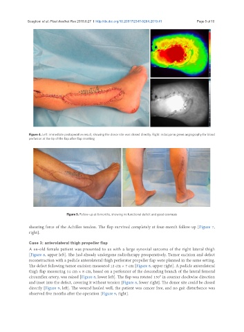
Figure 4. Left: immediate postoperative result, showing the donor site was closed directly. Right: indocyanin green angiography for blood
perfusion at the tip of the flap after flap insetting
Figure 5. Follow-up at 6 months, showing no functional deficit and good cosmesis
shearing force of the Achilles tendon. The flap survived completely at four-month follow-up [Figure 7,
right].
Case 3: anterolateral thigh propeller flap
A 66-old female patient was presented to us with a large synovial sarcoma of the right lateral thigh
[Figure 8, upper left]. She had already undergone radiotherapy preoperatively. Tumor excision and defect
reconstruction with a pedicle anterolateral thigh perforator propeller flap were planned in the same setting.
The defect following tumor excision measured 12 cm × 7 cm [Figure 8, upper right]. A pedicle anterolateral
thigh flap measuring 14 cm × 8 cm, based on a perforator of the descending branch of the lateral femoral
circumflex artery, was raised [Figure 8, lower left]. The flap was rotated 130° in counter clockwise direction
and inset into the defect, covering it without tension [Figure 8, lower right]. The donor site could be closed
directly [Figure 9, left]. The wound healed well, the patient was cancer free, and no gait disturbance was
observed five months after the operation [Figure 9, right].