
Page 71 - Read Online
P. 71
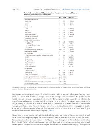
Page 8 of 11                                       Diamond et al. Plast Aesthet Res 2019;6:20  I  http://dx.doi.org/10.20517/2347-9264.2019.26

                             Table 4. Characteristics of 20 patients who underwent perforator based flaps for
                             treatment of lower extremity osteomyelitis
                                                                      No.  (%)   Clearance a
                              Soft tissue defect location
                                 Calf/Knee                            7    35    7 (100%)
                                 Ankle                                5    25    4 (80%)
                                 Foot                                 8    40    7 (87.5%)
                              Bone involved
                                 Tibia                                5    25    5 (100%)
                                 Fibula                               2    10    2 (100%)
                                 Calcaneus                            5    25    4 (80%)
                                 Ankle mortis/carpus                  6    30    6 (100%)
                                 Metatarsal/phalangeal                2    10    1 (50%)
                              Tissue based diagnosis                  20   100
                              Cierney-Mader classification
                                  I - Superficial                     12   60    12 (100%)
                                  II - Medullary                      3    15    2 (67%)
                                  III - Isolated (Sequestrum)         1    5     1 (100%)
                                  IV - Diffuse                        4    20    1 (75%)
                              Hardware present and kept in place      (4/4)  100  4 (100%)
                              External fixator present                2    10    2 (100%)
                              Microorganism
                                 Staph epidermidus, coagulase negative staph. MSSA  13  65  12 (92%)
                                 MRSA                                 1    5     1 (100%)
                                 Enterobacter                         2    10    2 (100%)
                                 Streptococcal                        1    5     1 (100%)
                                 Corynebacterium                      1    5     1 (100%)
                                 Proteus sp.                          1    5     0 (0%)
                                 Stenotrophomonas                     1    5     1 (100%)
                              Flap thickness
                                 Subfascial (Thick)                   4    20    4 (100%)
                                 Suprafascial                         6    30    5 (83%)
                                 Superthin (Periscarpal)              10   50    9 (90%)
                              Bone union achieved across fracture line  (8/9)  89%
                              External fixator exchanged for internal hardware or removed  (2/2)  100%
                              Amputation                              2    10
                              Radiographically healed compared to preoperative   (7/9)  78%
                              Osteomyelitis recurrence                2    10%

               a Osteomyelitis clearance as defined by lack of local recurrence of boney osteomyelitis, discontinuation of antibiotic, healed soft-tissue
               envelope, clearance of deep-space infection

               In subgroup analysis of our highest risk populations nine diabetic patients had osteomyelitis and three
               more had critical limb ischemia. Although this is a small sample size, outcomes in this population are
               mixed: none experienced recurrence of osteomyelitis defined as clinical evidence of bone infection by
               clinical exam, radiography or tissue pathology within the surgical site; five of our patients were fully
               weight-bearing in less than four months while three of them never fully ambulated due to conservative
               management of secondary ulcers in the same extremity, and one patient went on to amputation due to
               severe peripheral artery disease. Only one flap loss occurred due to extensive arterial thrombosis despite
               early intervention within the osteomyelitis group [Table 3].


               Microvascular tissue transfer in high-risk individuals harboring vascular disease, osteomyelitis and
               the Charcot foot improves upon outcomes achieved with alternative standard of care pathways.
               Revascularization alone as demonstrated in the “Bypass versus Angioplasty in Severe Ischemia of the Leg
                                [23]
               Trial” (BASIL Trial)  offers limited salvage rates with shortened up overall amputation-free survival and
               mortality when compared to revascularization plus wound directed reconstructive surgery. Of 250 patients