
Page 66 - Read Online
P. 66
Diamond et al. Plast Aesthet Res 2019;6:20 I http://dx.doi.org/10.20517/2347-9264.2019.26 Page 3 of 11
A B
C D
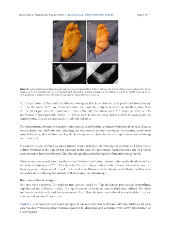
Figure 1. Superficial osteomyelitis managed with suprafascial anterolateral thigh. A patient with Cierny-Mader Class 2 ostomyelitis of the
calcaneus. A: A preoperative photo; B: immediate post flap phot; C: pre flap radiography with osteomyelitis; D: six-month follow up photo
with clearance of osteomyelitis. The patient was weight bearing at time of follow up
For the purposes of this study, the database was queried in June 2018 for cases performed from January
2015 to December 2017. We excluded muscle flaps and skin-only or fasciocutaneous flaps other than
ALT’s. Of 84 patients who underwent lower extremity free-tissue with ALT flaps, we excluded 25
individuals without high risk factors. This left 59 patients selected for at least one of the following features:
osteomyelitis, Charcot collapse, and critical limb ischemia.
For each patient, relevant demographic information, comorbidities, presence of peripheral vascular disease,
revascularization, antibiotic use, anticoagulant use, wound etiology, pre-operative imaging, anatomical
wound location, skeletal fixation, flap thickness, operative characteristics, complications and follow-up
were reviewed.
Osteomyelitis was defined as tissue-proven boney infection via histological analysis and bony tissue
culture obtained at the time of flap coverage in the case of single-staged reconstructions and or prior to
reconstruction from bone biopsy. Clinical, radiographic, microbiological information was gathered.
Patients were separated based on the Cierny-Mader classification system defining the depth as well as
diffusion of osteomyelitis [8,9,20] . Patients with Charcot collapse, critical limb ischemia (defined by vascular
imaging proven: single vessel run-off, multi-level or multivessel arterial disease) and diabetes mellitus were
separated into a subgroup for analysis of their unique pathophysiology.
Reconstructive technique
Patients were separated for analysis into groups based on flap thickness: periscarpal (superthin),
suprafascial and subfascial (thick). During the period of study, no muscle-flaps were utilized. We relied
uniformly on skin-only and fasciocutaneous flaps. Flap thickness was tailored to match defect surface
contour and volume of dead space.
Figures 1-3 demonstrate case based examples of our reconstructive technique. ALT flap thickness for each
case was determined by defect thickness, need to fill deadspace and correlates with Cierny classification of
Osteomyelitis.