
Page 67 - Read Online
P. 67
Page 4 of 11 Diamond et al. Plast Aesthet Res 2019;6:20 I http://dx.doi.org/10.20517/2347-9264.2019.26
A B
C D
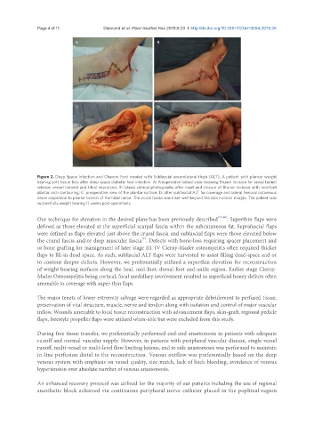
Figure 2. Deep Space Infection and Charcot Foot treated with Subfascial anterolateral thigh (ALT). A patient with plantar weight
bearing soft tissue loss after deep space diabetic foot infection. A: Preoperative lateral view showing Bruner incision for tarsal tunnel
release, vessel harvest and tibial neurolysis; B: lateral clinical photography after inset and closure of Bruner incision with mid-foot
plantar arch contouring; C: preoperative view of the plantar surface; D: after subfascial ALT for coverage and lateral femoral cutaneous
nerve coaptation to plantar branch of the tibial nerve. The crural fascia was inset well beyond the skin incision margin. The patient was
successfully weight bearing 11 weeks post-operatively
Our technique for elevation in the desired plane has been previously described [15,16] . Superthin flaps were
defined as those elevated at the superficial scarpal fascia within the subcutaneous fat. Suprafascial flaps
were defined as flaps elevated just above the crural fascia and subfascial flaps were those elevated below
[2]
the crural fascia and/or deep muscular fascia . Defects with bone-loss requiring spacer placement and
or bone grafting for management of later stage III, IV Cierny-Mader osteomyelitis often required thicker
flaps to fill-in dead space. As such, subfascial ALT flaps were harvested to assist filling dead-space and or
to contour deeper defects. However, we preferentially utilized a superthin elevation for reconstruction
of weight bearing surfaces along the heal, mid-foot, dorsal-foot and ankle region. Earlier stage Cierny-
Mader Osteomyelitis being cortical, focal medullary involvement resulted in superficial boney defects often
amenable to coverage with super-thin flaps.
The major tenets of lower extremity salvage were regarded as appropriate debridement to perfused tissue,
preservation of vital structure, muscle, nerve and tendon along with isolation and control of major vascular
inflow. Wounds amenable to local tissue reconstruction with advancement flaps, skin-graft, regional pedicle
flaps, freestyle propeller flaps were utilized when-able but were excluded from this study.
During free tissue transfer, we preferentially performed end-end anastomosis in patients with adequate
runoff and normal vascular supply. However, in patients with peripheral vascular disease, single-vessel
runoff, multi-vessel or multi-level flow limiting lesions, end to side anastomosis was performed to maintain
in-line perfusion distal to the reconstruction. Venous outflow was preferentially based on the deep
venous system with emphasis on vessel quality, size-match, lack of back-bleeding, avoidance of venous
hypertension over absolute number of venous anastomosis.
An enhanced recovery protocol was utilized for the majority of our patients including the use of regional
anesthetic block achieved via continuous peripheral nerve catheter placed in the popliteal region