
Page 35 - Read Online
P. 35
Page 4 of 9 Schuster. Plast Aesthet Res 2018;5:22 I http://dx.doi.org/10.20517/2347-9264.2018.13
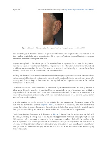
Figure 3. Skin erosion of the upper edge of an implant placed over the superior crus of the antihelical fold
dure. Interestingly, of those who declined to go ahead with treatment using Earfold™, none went on to opt
for a standard surgical alternative suggesting that this is a group of patients who would not otherwise come
forward for treatment of their prominent ears.
Implants were placed at the inferior part of the antihelical fold in 5 patients. In 19 cases, the implant was
placed at the superior part of the antihelical fold (11 proximal to the bifurcation, 8 distal to the bifurcation).
In addition, surgery to reduce the size of the anti-tragus was performed bilaterally in 1 patient. In further 4
patients, Earfold™ was used in combination with Mustardé sutures.
Building familiarity with the introducer in the weeks before surgery is performed is critical for eventual cor-
rect deployment of the implant. In 2 cases, the implants had to be discarded as the implants were noted to be
sitting proud of the cartilage. In those cases, the cartilage had not been kept flat in relation to the cartilage
during release of the implant.
The author did not use a validated method of assessment of patient satisfaction and the average duration of
follow-up in this series was short (6 months). However, anecdotally, 16 out of 19 patients were satisfied or
very satisfied with the aesthetic result. Three patients were dissatisfied with the outcome of treatment due to
issues with persistent pain and sensitivity, which were resolved after removal of the implants and the perfor-
mance of a Mustardé otoplasty.
In total, the author removed 8 implants from 5 patients. Removal was necessary because of erosion of the
skin over the implant in 2 patients [Figures 3 and 4] and because of continuing pain and inflammation
around the implant in 2 cases. In one case, the positioning of the implant was aesthetically unsatisfying, in-
ducing an excessive folding. Removals were performed within 2 to 15 months after implantation.
Careful examination of the cases with skin erosion [Figure 3] showed that the implant was not flush with
the cartilage resulting in a sharp edge of the implant sitting proud and eventually eroding through the skin.
Although every effort was made to ensure that the implants were completely flush with the cartilage at the
time of deployment, it is entirely possible that minor mispositioning of the implant was not detected due to
soft-tissue swelling at the time of surgery. Alternatively, the implants may have moved due to patient factors
during the first 3-4 weeks after surgery (i.e., before encapsulation of the implant occurred preventing any
further movement).