
Page 36 - Read Online
P. 36
Schuster. Plast Aesthet Res 2018;5:22 I http://dx.doi.org/10.20517/2347-9264.2018.13 Page 5 of 9
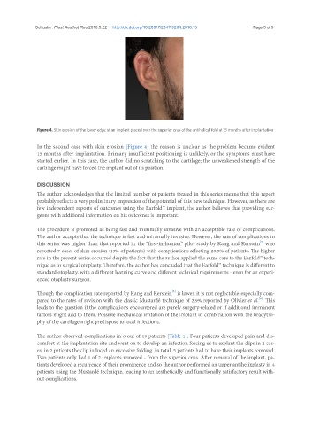
Figure 4. Skin erosion of the lower edge of an implant placed over the superior crus of the antihelical fold at 15 months after implantation
In the second case with skin erosion [Figure 4] the reason is unclear as the problem became evident
15 months after implantation. Primary insufficient positioning is unlikely, or the symptoms must have
started earlier. In this case, the author did no scratching to the cartilage; the unweakened strength of the
cartilage might have forced the implant out of its position.
DISCUSSION
The author acknowledges that the limited number of patients treated in this series means that this report
probably reflects a very preliminary impression of the potential of this new technique. However, as there are
few independent reports of outcomes using the Earfold™ implant, the author believes that providing sur-
geons with additional information on his outcomes is important.
The procedure is promoted as being fast and minimally invasive with an acceptable rate of complications.
The author accepts that the technique is fast and minimally invasive. However, the rate of complications in
[3]
this series was higher than that reported in the “first-in-human” pilot study by Kang and Kerstein who
reported 7 cases of skin erosion (13% of patients) with complications affecting 20.5% of patients. The higher
rate in the present series occurred despite the fact that the author applied the same care to the Earfold™ tech-
nique as to surgical otoplasty. Therefore, the author has concluded that the Earfold™ technique is different to
standard otoplasty, with a different learning curve and different technical requirements - even for an experi-
enced otoplasty surgeon.
[3]
Though the complication rate reported by Kang and Kerstein is lower, it is not neglectable-especially com-
[6]
pared to the rates of revision with the classic Mustardé technique of 2.9% reported by Olivier et al. . This
leads to the question if the complications encountered are purely surgery-related or if additional immanent
factors might add to them. Possible mechanical irritation of the implant in combination with the bradytro-
phy of the cartilage might predispose to local infections.
The author observed complications in 6 out of 19 patients [Table 1]. Four patients developed pain and dis-
comfort at the implantation site and went on to develop an infection forcing us to explant the clips in 2 cas-
es; in 2 patients the clip induced an excessive folding. In total, 5 patients had to have their implants removed.
Two patients only had 1 of 2 implants removed - from the superior crus. After removal of the implant, pa-
tients developed a recurrence of their prominence and so the author performed an upper antihelixplasty in 4
patients using the Mustardé technique, leading to an aesthetically and functionally satisfactory result with-
out complications.