
Page 37 - Read Online
P. 37
Page 6 of 9 Schuster. Plast Aesthet Res 2018;5:22 I http://dx.doi.org/10.20517/2347-9264.2018.13
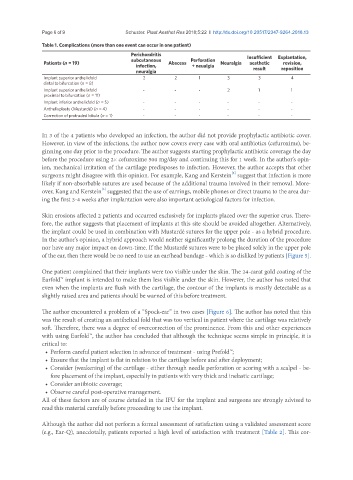
Table 1. Complications (more than one event can occur in one patient)
Perichondritis Insufficient Explantation,
subcutaneous Perforation
Patients (n = 19) Abscess Neuralgia aesthetic revision,
infection, + neualgia result reposition
neuralgia
Implant superior anthelixfold 2 2 1 3 3 4
distal to bifurcation (n = 8)
Implant superior anthelixfold - - - 2 1 1
proximal to bifurcation (n = 11)
Implant inferior anthelixfold (n = 5) - - - - - -
Anthelixplasty (Mustardé) (n = 4) - - - - - -
Correction of protruded lobula (n = 1) - - - - - -
In 3 of the 4 patients who developed an infection, the author did not provide prophylactic antibiotic cover.
However, in view of the infections, the author now covers every case with oral antibiotics (cefuroxime), be-
ginning one day prior to the procedure. The author suggests starting prophylactic antibiotic coverage the day
before the procedure using 2× cefuroxime 500 mg/day and continuing this for 1 week. In the author’s opin-
ion, mechanical irritation of the cartilage predisposes to infection. However, the author accepts that other
surgeons might disagree with this opinion. For example, Kang and Kerstein suggest that infection is more
[3]
likely if non-absorbable sutures are used because of the additional trauma involved in their removal. More-
over, Kang and Kerstein suggested that the use of earrings, mobile phones or direct trauma to the area dur-
[3]
ing the first 3-4 weeks after implantation were also important aetiological factors for infection.
Skin erosions affected 2 patients and occurred exclusively for implants placed over the superior crus. There-
fore, the author suggests that placement of implants at this site should be avoided altogether. Alternatively,
the implant could be used in combination with Mustardé sutures for the upper pole - as a hybrid procedure.
In the author’s opinion, a hybrid approach would neither significantly prolong the duration of the procedure
nor have any major impact on down-time. If the Mustardé sutures were to be placed solely in the upper pole
of the ear, then there would be no need to use an ear/head bandage - which is so disliked by patients [Figure 5].
One patient complained that their implants were too visible under the skin. The 24-carat gold coating of the
Earfold™ implant is intended to make them less visible under the skin. However, the author has noted that
even when the implants are flush with the cartilage, the contour of the implants is mostly detectable as a
slightly raised area and patients should be warned of this before treatment.
The author encountered a problem of a “Spock-ear” in two cases [Figure 6]. The author has noted that this
was the result of creating an antihelical fold that was too vertical in patient where the cartilage was relatively
soft. Therefore, there was a degree of overcorrection of the prominence. From this and other experiences
with using Earfold™, the author has concluded that although the technique seems simple in principle, it is
critical to:
• Perform careful patient selection in advance of treatment - using Prefold™;
• Ensure that the implant is flat in relation to the cartilage before and after deployment;
• Consider (weakening) of the cartilage - either through needle perforation or scoring with a scalpel - be-
fore placement of the implant, especially in patients with very thick and inelastic cartilage;
• Consider antibiotic coverage;
• Observe careful post-operative management.
All of these factors are of course detailed in the IFU for the implant and surgeons are strongly advised to
read this material carefully before proceeding to use the implant.
Although the author did not perform a formal assessment of satisfaction using a validated assessment score
(e.g., Ear-Q), anecdotally, patients reported a high level of satisfaction with treatment [Table 2]. This cor-