
Page 40 - Read Online
P. 40
Page 8 of 17 Garoosi et al. Plast Aesthet Res 2024;11:42 https://dx.doi.org/10.20517/2347-9264.2024.57
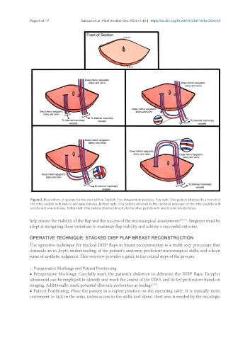
Figure 2. Illustrations of options for the stacked flap.Top left: Two independent pedicles. Top right: One pedicle attached to a branch of
the other pedicle with end-to-end anastomoses. Bottom right: One pedicle attached to the cephalad extension of the other pedicle with
end-to-end anastomoses. Bottom left: One pedicle attached directly to the other pedicle with end-to-side anastomoses.
help ensure the viability of the flap and the success of the microsurgical anastomoses [24-26] . Surgeons must be
adept at navigating these variations to maximize flap viability and achieve a successful outcome.
OPERATIVE TECHNIQUE: STACKED DIEP FLAP BREAST RECONSTRUCTION
The operative technique for stacked DIEP flaps in breast reconstruction is a multi-step procedure that
demands an in-depth understanding of the patient’s anatomy, proficient microsurgical skills, and a keen
sense of aesthetic judgment. This overview provides a guide to the critical steps of the process.
1. Preoperative Markings and Patient Positioning
● Preoperative Markings: Carefully mark the patient’s abdomen to delineate the DIEP flaps. Doppler
ultrasound can be employed to identify and mark the course of the DIEA and its key perforators based on
imaging. Additionally, mark potential alternate perforators as backup [1,18] .
● Patient Positioning: Place the patient in a supine position on the operating table. It is typically more
convenient to tuck in the arms, unless access to the axilla and lateral chest area is needed by the oncologic