
Page 37 - Read Online
P. 37
Garoosi et al. Plast Aesthet Res 2024;11:42 https://dx.doi.org/10.20517/2347-9264.2024.57 Page 5 of 17
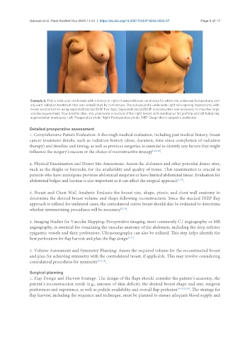
Example 2. This is a 66-year-old female with a history of right intraductal breast carcinoma for which she underwent lumpectomy and
adjuvant radiation treatment that was complicated by contracture. She subsequently underwent right skin-sparing mastectomy with
breast reconstruction using separated stacked DIEP free flaps. Separated stacked DIEP reconstruction was necessary to meet her large
volume requirement. Four months later, she underwent a revision of her right breast with autologous fat grafting and left balancing
augmentation mastopexy. Left: Preoperative photo. Right: Postoperative photo. DIEP: Deep inferior epigastric perforator.
Detailed preoperative assessment
1. Comprehensive Patient Evaluation: A thorough medical evaluation, including past medical history, breast
cancer treatment details, such as radiation history (dose, duration, time since completion of radiation
therapy) and timeline and timing, as well as previous surgeries, is essential to identify any factors that might
influence the surgery’s success or the choice of reconstructive strategy [1,16,18] .
2. Physical Examination and Donor Site Assessment: Assess the abdomen and other potential donor sites,
such as the thighs or buttocks, for the availability and quality of tissue. This examination is crucial in
patients who have undergone previous abdominal surgeries or have limited abdominal tissue. Evaluation for
abdominal bulges and hernias is also important as it can affect the surgical approach [1,18] .
3. Breast and Chest Wall Analysis: Evaluate the breast size, shape, ptosis, and chest wall anatomy to
determine the desired breast volume and shape following reconstruction. Since the stacked DIEP flap
approach is utilized for unilateral cases, the contralateral native breast should also be evaluated to determine
whether symmetrizing procedures will be necessary [13,18] .
4. Imaging Studies for Vascular Mapping: Preoperative imaging, most commonly CT angiography or MR
angiography, is essential for visualizing the vascular anatomy of the abdomen, including the deep inferior
epigastric vessels and their perforators. Ultrasonography can also be utilized. This step helps identify the
best perforators for flap harvest and plan the flap design [1,19] .
5. Volume Assessment and Symmetry Planning: Assess the required volume for the reconstructed breast
and plan for achieving symmetry with the contralateral breast, if applicable. This may involve considering
contralateral procedures for symmetry [1,10,19] .
Surgical planning
1. Flap Design and Harvest Strategy: The design of the flaps should consider the patient’s anatomy, the
patient’s reconstruction needs (e.g., amount of skin deficit), the desired breast shape and size, surgeon
preferences and experience, as well as pedicle availability and overall flap perfusion [10,18,22,23] . The strategy for
flap harvest, including the sequence and technique, must be planned to ensure adequate blood supply and