
Page 38 - Read Online
P. 38
Page 6 of 17 Garoosi et al. Plast Aesthet Res 2024;11:42 https://dx.doi.org/10.20517/2347-9264.2024.57
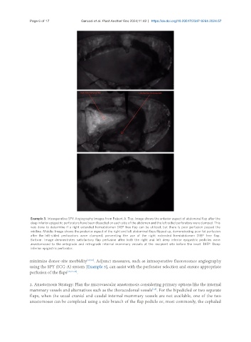
Example 3. Intraoperative SPY Angiography Images from Patient A. Top: Image shows the anterior aspect of abdominal flap after the
deep inferior epigastric perforators have been dissected on each side of the abdomen and the left-sided perforators were clamped. This
was done to determine if a right extended hemiabdomen DIEP free flap can be utilized, but there is poor perfusion passed the
midline. Middle: Image shows the posterior aspect of the right and left abdominal flaps flipped up, demonstrating poor fat perfusion
after the left-sided perforators were clamped, preventing the use of the right extended hemiabdomen DIEP free flap.
Bottom: Image demonstrates satisfactory flap perfusion after both the right and left deep inferior epigastric pedicles were
anastomosed to the antegrade and retrograde internal mammary vessels at the recipient site before the inset. DIEP: Deep
inferior epigastric perforator.
minimize donor-site morbidity [1,5,10] . Adjunct measures, such as intraoperative fluorescence angiography
using the SPY (ICG-A) system [Example 3], can assist with the perforator selection and ensure appropriate
perfusion of the flaps [10,24-26] .
2. Anastomosis Strategy: Plan the microvascular anastomosis considering primary options like the internal
[1,5]
mammary vessels and alternatives such as the thoracodorsal vessels . For the bipedicled or two separate
flaps, when the usual cranial and caudal internal mammary vessels are not available, one of the two
anastomoses can be completed using a side branch of the flap pedicle or, most commonly, the cephalad