
Page 65 - Read Online
P. 65
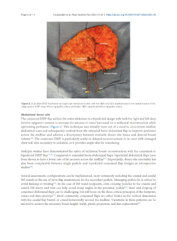
Page 4 of 11          Farajzadeh et al. Plast Aesthet Res 2024;11:32  https://dx.doi.org/10.20517/2347-9264.2024.24


                Figure 2. Dual-plane DIEP flap based on single high medial perforator with the SIEA and SIEV anastomosed to the lateral branch of the
                deep system. DIEP: deep inferior epigastric artery perforator; SIEA: superficial inferior epigastric artery.


               Abdominal donor site
               The conjoined DIEP flap utilizes the entire abdomen in a bipedicled design with both the right and left deep
               inferior epigastric systems to increase the amount of tissue harvested for a unilateral reconstruction while
               optimizing perfusion [Figure 3]. This technique was initially born out of a need to circumvent midline
               abdominal scars and subsequently evolved from the extended hemi-abdominal flap to improve perfusion
               across the midline and address a discrepancy between available donor-site tissue and desired breast
                     [14]
               volume . The conjoined DIEP is particularly useful in delayed reconstructions or in cases with damaged
               chest wall skin secondary to radiation, as it provides ample skin for resurfacing.


               Multiple studies have demonstrated the safety of unilateral breast reconstruction with the conjoined or
               bipedicled DIEP flap [15-20] . Compared to extended hemi-abdominal flaps, bipedicled abdominal flaps have
               been shown to have a lower rate of fat necrosis across the midline . Importantly, donor-site morbidity has
                                                                       [21]
               also been comparable between single pedicle and bipedicled conjoined flap designs in retrospective
               studies .
                     [22]

               Several anastomotic configurations can be implemented, most commonly including the cranial and caudal
               IM vessels or the use of intra-flap anastomoses for the secondary pedicle. Managing pedicle lie is critical to
               avoid kinking or twisting . In the case of IM vessel recipients, criss-crossing pedicles to the cranial and
                                     [23]
                                                                                     [21]
               caudal IM artery and vein can help avoid sharp angles in the proximal pedicle . Inset and shaping of
               conjoined abdominal flaps can be challenging, but still focus on the three critical principles of the footprint,
                                    [24]
               conus and skin envelope . Most commonly, conjoined flaps are either folded in the vertical dimension
               with the caudal flap buried or coned horizontally around the midline. Variations in these patterns can be
                                                                                              [25]
               tailored to achieve the necessary breast height, width, ptosis, projection, and skin replacement .