
Page 69 - Read Online
P. 69
Page 8 of 11 Farajzadeh et al. Plast Aesthet Res 2024;11:32 https://dx.doi.org/10.20517/2347-9264.2024.24
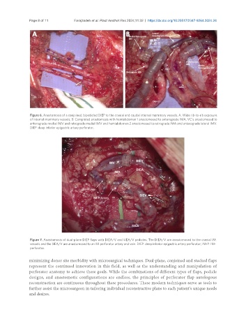
Figure 6. Anastomosis of a conjoined, bipedicled DIEP to the cranial and caudal internal mammary vessels. A: Wide rib-to-rib exposure
of internal mammary vessels; B: Completed anastomosis with hemiabdomen 1 anastomosed to anterograde IMA, VC’s anastomosed to
anterograde medial IMV and retrograde medial IMV and hemiabdomen 2 anastomosed to retrograde IMA and anterograde lateral IMV.
DIEP: deep inferior epigastric artery perforator.
Figure 7. Anastomosis of dual-plane DIEP flaps with DIEA/V and SIEA/V pedicles. The DIEA/V are anastomosed to the cranial IM
vessels and the SIEA/V are anastomosed to an IM perforator artery and vein. DIEP: deep inferior epigastric artery perforator; IM-P: IM-
perforator.
minimizing donor site morbidity with microsurgical techniques. Dual-plane, conjoined and stacked flaps
represent the continued innovation in this field, as well as the understanding and manipulation of
perforator anatomy to achieve these goals. While the combinations of different types of flaps, pedicle
designs, and anastomotic configurations are endless, the principles of perforator flap autologous
reconstruction are continuous throughout these procedures. These modern techniques serve as tools to
further assist the microsurgeon in tailoring individual reconstructive plans to each patient’s unique needs
and desires.