
Page 10 - Read Online
P. 10
Sarrami et al. Plast Aesthet Res 2024;11:13 https://dx.doi.org/10.20517/2347-9264.2024.06 Page 5 of 15
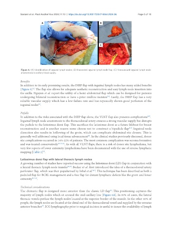
Figure 4. (A) Identification of inguinal lymph nodes; (B) Harvested inguinal lymph node flap; (C) Vascularized inguinal lymph node
anastomosis to axillary blood supply.
Benefits
In addition to its early promising results, the DIEP flap with inguinal lymph nodes has many added benefits
[1]
[Figure 5] . The flap size allows for adequate aesthetic reconstruction and easy lymph node insertion into
the axilla. Nguyen et al. report the utility of a hemi-abdominal flap which can be designed for patients
[16]
undergoing bilateral reconstruction or have a prior midline incision . Lastly, the DIEP flap has a very
reliable vascular supply which has a low failure rate and has repeatedly shown good perfusion of the
inguinal nodes .
[8]
Pitfalls
In addition to the risks associated with the DIEP flap alone, the VLNT flap also presents complications .
[27]
Inguinal lymph node anastomosis to the thoracodorsal artery ensures a strong vascular supply but disrupts
the pedicle to the latissimus dorsi flap. This sacrifices the latissimus dorsi as a future lifeboat for breast
[5,7]
reconstruction and is another reason some choose not to construct a bipedicle flap . Inguinal node
dissection also results in hollowing of the groin, which can complicate abdominal site closure. This is
generally well addressed using local tissue advancement . In the clinical studies previously discussed, donor
[8]
site complications occurred in 14%-22% of patients. The most common complication was seroma formation
and was treated conservatively [16,25,26] . As with all VLNT flaps, there is a risk of donor site lymphedema, but
very few reports of lower extremity lymphedema have been documented with the use of reverse lymphatic
mapping [Table 2] .
[5]
Latissimus dorsi flap with lateral thoracic lymph nodes
A growing number of studies have reported success using the latissimus dorsi (LD) flap in conjunction with
a lateral thoracic lymph node transfer [4,28] . Becker et al. first introduced the idea of a thoracodorsal artery
perforator flap, which was then popularized by Inbal et al. . This technique has been described as both a
[4,7]
pedicled flap for BCRL management and a free flap for distant lymphatic defects like the groin and lower
extremity [5,7,11] .
Technical conisderations
The chimeric flap is designed more anterior than the classic LD flap . This positioning captures the
[4]
majority of lymph nodes which sit around the mid-axillary line [Figure 6A]. In 60% of cases, the lateral
thoracic vessels perfuse the lymph nodes located at the superior border of the muscle. In the other 40% of
people, the lymph nodes are located at the distal end of the thoracodorsal vessel and supplied by the serratus
anterior branches . ICG lymphography prior to surgical incision is useful to insure the availability of lymph
[7]