
Page 14 - Read Online
P. 14
Sarrami et al. Plast Aesthet Res 2024;11:13 https://dx.doi.org/10.20517/2347-9264.2024.06 Page 9 of 15
Figure 9. Preoperative planning of SCIP flap with inguinal lymph node harvest (top). Dissection of flap perforator and inguinal lymph
nodes (bottom). SCIP: superficial circumflex iliac perforator.
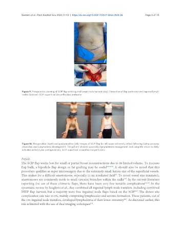
Figure 10. Preoperative (right) and postoperative (left) images of SCIP flap for left upper extremity defect following Ewing sarcoma
dissection and lymphedema development. The patient showed successful lymphedema management and complete return to daily
activities within a year postoperatively. SCIP: superficial circumflex iliac perforator.
Pitfalls
The SCIP flap works best for small or partial breast reconstructions due to its limited volume. To increase
flap bulk, a bipedicle flap design or fat grafting may be useful [27,30,36] . It should also be noted that this
procedure qualifies as super microsurgery due to the extremely small lumen size of the superficial vessels.
This makes for a difficult anastomosis, especially in an irradiated field . To avoid vessel size mismatch,
[5]
[33]
anastomoses are commonly made to small vascular branches within the axilla . In the current literature
reporting the use of these chimeric flaps, there have been very few notable complications [26,32] . In the
systematic review by Scaglioni et al., they combined all inguinal lymph node transfers, including combined
DIEP flap harvest, but a majority were free inguinal node flaps based on the SCIP . The donor site
[29]
complication rate was 10.9%, mainly comprising lymphoceles and seroma formation. Three patients, out of
the 195 inguinal node transfers, developed lymphedema of their lower extremity . As discussed earlier, this
[29]
[18]
risk is limited with the use of dual imaging techniques .