
Page 9 - Read Online
P. 9
Page 4 of 13 Bansberg et al. Plast Aesthet Res 2024;11:12 https://dx.doi.org/10.20517/2347-9264.2023.109
Table 1. Patient demographics, perforation size, surgical vs. non-surgical etiology
Patient (n) 392
Gender
Female n (%) 245 (62.5%)
Male n (%) 147 (37.5%)
Mean age years (range) 49.2 (14-81)
Perforation size
Mean length mm (range) 14.1 (2-37)
Mean height mm (range) 10.5 (2-20)
Perforation etiology
Surgical n (%) 159 (40.6%)
Non-surgical n (%) 233 (59.4%)
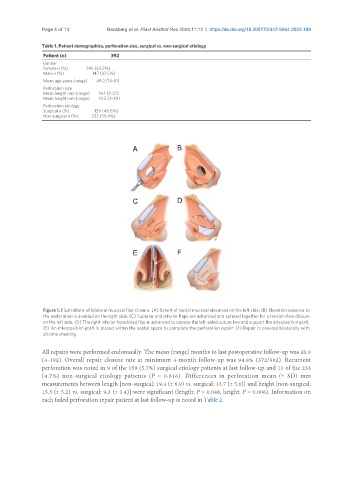
Figure 1. Illustrations of bilateral mucosal flap closure. (A) Extent of septal mucosal elevation on the left side; (B) Elevation superior to
the perforation is avoided on the right side; (C) Superior and inferior flaps are advanced and sutured together for a tension-free closure
on the left side; (D) The right inferior bipedicled flap is advanced to oppose the left-sided suture line and support the interposition graft;
(E) An interposition graft is placed within the septal space to complete the perforation repair; (F) Repair is covered bilaterally with
silicone sheeting.
All repairs were performed endonasally. The mean (range) months to last postoperative follow-up was 20.9
(4-192). Overall repair closure rate at minimum 4-month follow-up was 94.8% (372/392). Recurrent
perforation was noted in 9 of the 159 (5.7%) surgical etiology patients at last follow-up and 11 of the 233
(4.7%) non-surgical etiology patients (P = 0.816). Differences in perforation mean (± SD) mm
measurements between length [non-surgical: 19.4 (± 6.9) vs. surgical: 13.7 (± 5.0)] and height [non-surgical:
15.5 (± 5.2) vs. surgical: 9.3 (± 3.4)] were significant (length: P = 0.048, height: P = 0.006). Information on
each failed perforation repair patient at last follow-up is noted in Table 2.