
Page 11 - Read Online
P. 11
Page 6 of 13 Bansberg et al. Plast Aesthet Res 2024;11:12 https://dx.doi.org/10.20517/2347-9264.2023.109
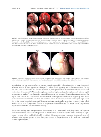
Figure 2. Endoscopic photos of left-side mucosal flap closure. (A) Perforation margin rimmed with #15 blade; (B) Bilateral mucosal flap
elevation; (C) Superior incision may extend anteriorly through valve angle; (D) Superior flap advanced inferiorly; (E) Anterior cautery
incision for inferior flap; (F) Elevation connecting incision over piriform aperture to previously elevated nasal floor mucosa; (G) Inferior
flap incision made with scissors; (H) Flaps advanced to collapse perforation margin for tension-free suture closure; Flaps span (bridge)
over the underlying defect’s cartilage margin.
Figure 3. Endoscopic pictures of 1.4 × 1.1 cm perforation repair utilizing left superior and inferior flaps, and right inferior flap, following
silicone sheeting removal at 12 days postoperative. Note the difference in repair thickness between the right and left sides.
visualization can improve septal space surgical precision, especially when attempting to separate scarred,
[14]
adherent mucosa following prior septal surgery . Bilateral and opposing mucosal tears that occur during
mucosal elevation increase the risk for perforation, though unilateral tears have been associated with
[15]
perforation development . We routinely place a mosaic of septal cartilage/bone pieces back into the septal
space at the procedure’s conclusion for mucosal flap and airway support. Thin rigid splints are applied for
septal stabilization and to minimize hematoma risk. In the presence of bilateral opposing tears, these
maneuvers serve to support a unilateral mucosal tear repair. A soft tissue acellular dermal graft is placed in
the septal space opposite the repair if bone or cartilage is not available for this purpose. Septal splint
application for 7-10 days prevents desiccation to promote mucosal healing. The senior author’s septoplasty
experience contributed one patient to this study.
Perforation etiology is not always apparent. Patients may have a history with multiple potential etiologies, or
deny any condition associated with perforation development. Perforation patients with a history of septal
surgery present with a readily identifiable event that determines etiology which may be clinically relevant
when considering management options. Forty-one percent of the perforations in this study were attributed
to prior septal surgery.